Automated imaging and detection of cytolytic plaques for neutralization assay
- A monolayer of host cells are seeded into a 6-, 12-, or 24-well plate
- Mixtures of the virus and various dilutions of the antibodies are produced and allowed to incubate for 2 hours
- After incubation, the different mixtures are then added to the host cells and incubated for another 2 hours
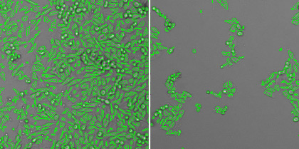
- The results showed a dose dependent decrease in the formation of cytolytic plaques as the antibody concentration increased
- In addition, plaque number and size information can be directly exported into excel for additional plotting and analysis

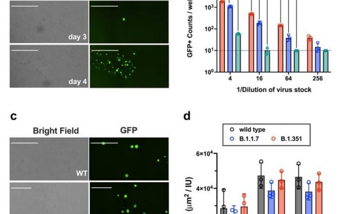
The Celigo Image Cytometer can directly count cytolytic plaques in plates

Plate-view of counted cytolytic plaques treated with different antibody concentrations in a 12-well plate
| Plaque count | |||
|---|---|---|---|
| 67 | 19 | 4 | 2 |
| 63 | 13 | 2 | 0 |
Plaque count and size measurement from the Celigo
| Plaque size (µm2) | |||
|---|---|---|---|
| 453017.801 | 478668.211 | 375579.65 | 58492.7559 |
| 481462.961 | 313941.745 | 382699.547 | NaN |
The Celigo Image Cytometer automates antibody neutralization assays:
Automated imaging and detection of cytolytic plaques
The Celigo Image Cytometry system performs high-througput, whole-well imaging and quantitative data in bright field and up to four fluorescent channels for a wide variety of cell-based assays.
Learn more about modern virology assays: