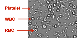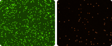
The viability and concentration of isolated PBMCs are traditionally measured by manual counting with trypan blue using a hemacytometer. One of the common issues of PBMC isolation is red blood cell (RBC) contamination. The RBC contamination can be dependent on the donor sample and/or technical skill level of the operator. RBC contamination in a PBMC sample can introduce error to the measured concentration, which can pass down to future experimental assays performed on these cells.
To resolve this issue, RBC lysing protocol can be used to eliminate potential error caused by RBC contamination. In recent years, a rapid fluorescence-based image cytometry system has been utilized for bright-field and fluorescence imaging analysis of cellular characteristics.
Nexcelom’s Cellometer image cytometry system has demonstrated the capability of automated concentration and viability detection in disposable counting chambers of unpurified mouse splenocytes and PBMCs stained with acridine orange and propidium idodide under fluorescence detection.
In a recently published paper, we demonstrate the ability of the Cellometer system to accurately measure PBMC concentration, despite RBC contamination, by comparison of five different total PBMC counting methods: manual counting of trypan blue-stained PBMCs in hemacytometer, manual counting of PBMCs in bright-field images, manual counting of acetic acid lysing of RBCs with TB-stained PBMCs, automated counting of acetic acid lysing of RBCs with PI-stained PBMCs, and AO/PI dual staining method.
The results show comparable total PBMC counting among all five methods, which validate the AO/PI staining method for PBMC measurement in the image cytometry method.
To read this paper in its entirety, please see J Immunol Methods. 2012 Nov 29. pii: S0022-1759(12)00343-2.


Leave A Comment