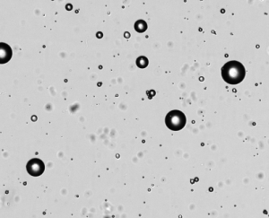
Adipocytes are the primary cells found in adipose (fat) tissue. Adipocytes store energy for the body in the form of triacylglycerol and play a critical role in lipid metabolism and energy regulation. Adipocytes have also been found to express a wide range of cytokines, chemokines, receptors, and cell adhesion molecules capable of inducing inflammation. Though it was once believed that mature adipocytes do not proliferate, a small degree of proliferation has been observed in mature adipocytes in culture. It is likely that overall adipocyte cell number is maintained over time by highly-controlled apoptosis and proliferation.
Obesity is associated with an increase in adipocyte size due to the accumulation of triacylglycerol (lipid) that can trigger changes in adipocyte hormone and cytokine secretion. The trigger of this inflammatory response from adipocytes is thought to lead to several obesity-related diseases including chronic inflammation, insulin resistance, type 2 diabetes, and cardiovascular disease.
Though the majority of adipose tissue in humans is white adipose tissue (WAT) involved with fat storage, a small amount of brown adipose tissue (BAT) exists in distinct locations within the body. The function of BAT is to burn fat for heat production. Researchers are exploring ways to induce the conversion of WAT to BAT to fight obesity and are investigating the differences in the origin and differentiation of adipocytes found in WAT and BAT.
When cultured, adipocytes float on top of the medium and clump together, limiting their access to nutrients in the medium. Most adipocytes undergo cell lysis within 72 hours of isolation. The buoyancy and fragility of adipocytes has led to the development of some unique culture methods. In some cases, researchers culture pre-adipocytes for differentiation into adipocytes. Some researchers have employed a ceiling culture method where isolated adipocytes adhere to a floating glass surface and remain viable for several weeks. Other researchers have utilized adipose tissue fragments embedded in a three-dimensional collagen gel that traps buoyant adipocytes. This method has been shown to maintain the normal function and proliferative ability of mature adipocytes for more than four weeks.
Clone Evaluated: primary adipocytes
Adherent / Non-adherent: predominantly adherent
Cell Type: mesenchymal cell
Cell Source: white adipose (fat) tissue
Cell Characteristics: Differentiated adipocytes are spherical in shape with differences in fat organelle (lipid droplet) size. Most space in the adipocyte is composed of fat organelles. Mature adipocytes often contain a single large lipid droplet. Adipocytes can be much larger than most mammalian cells, with a diameter of up to ~300 microns.
Recommended Media: Different media formulations are available for growth of pre-adipocytes, differentiation of pre-adipocytes into adipocytes, and growth of mature adipocytes.
Though most automated cell counters and flow cytometers cannot analyze adipocytes due to their size and/or fragility, Nexcelom has developed special large-depth cell counting chambers specifically for use with adipocytes and other large cells. In the Cellometer image cytometry method, 20µl of cell sample is pipetted into the counting chamber. Cells are imaged and analyzed directly from the counting chamber, enabling analysis of fragile cells, including adipocytes and hepatocytes. The Cellometer Vision Cell Analyzer and Cellometer Vision CBA Image Cytometry System are equipped with specialized settings for accurate determination of adipocyte concentration and viability.
Adipocyte References:
Meijer, K., et al. (2011). Human Primary Adipocytes Exhibit Immune Cell Function: Adipocytes Prime Inflammation Independent of Macrophages, PLoS ONE. 6(3), e17154. Doi: 10.1371/journal.pone.0017154.
Zhang, H. et al. (2000). Ceiling Culture of Mature Human Adipocytes: Use in Studies of Adipocyte Functions, Journal of Endocrinology. 164:119-128.
Sonodai, E. et al. (2008). A New Organotypic Culture of Adipose Tissue Fragments Maintains Viable Mature Adipocytes for a Long Term, Together with Development of Immature Adipocytes and Mesenchymal Stem Cell-Like Cells, Endochrinology. 149(10):4794-4798. Doi: 10.1210/en.2008-0525.
Farmer, S. (2008). Molecular Determinants of Brown Adipocyte Formation and Function. Genes and Development. 22: 1269-1275.


Leave A Comment