
Evaluating Anticancer Drug Compounds using Image Cytometry
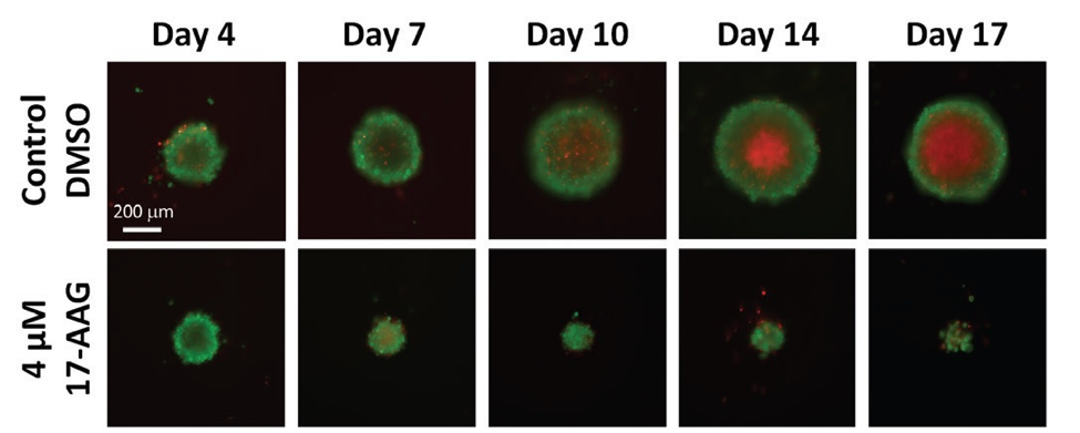
Image-based, automated analysis of 3-D tumor spheroids in microwell plates is becoming standard practice for the evaluation of anticancer drug compounds.
Image-based, automated analysis of 3-D tumor spheroids in microwell plates is becoming standard practice for the evaluation of anticancer drug compounds.
November is National Alzheimer's Disease Awareness Month and research into a cure for the disease is vigorous. In a recently published paper "CRISPR/Cas9-Correctable mutation-related molecular andphysiological phenotypes in iPSC-derived Alzheimer’s PSEN2N141I neurons" researchers used the Celigo image cytometer upstream of CRISPR/Cas9 to visualize and asses cell death related to the sensitivity of iPSC-derived PSEN2N1411 neurons in the presence of Aβ42 oligomer toxicity.
Acquire Fast, Accurate and Consistent Cell Viability and Concentration Measurements of Small Cells (2 microns or greater)! Obtaining consistent viability and concentration measurements for small cells like yeast, platelets or algae can be challenging. The Cellometers X1 and X2 can both perform bright-field and fluorescent counting of cells 2 microns and greater. X1 and X2 Cellometers are equipped with a 10X magnification lens. Both are capable of providing consistent concentration and viability measurements for cell samples that are 2 microns in size and greater. Both instruments are routinely used in laboratories around the world interested in studying small cells. See [...]
Obtain fast, accurate and consistent cell viability and concentration measurements of primary murine samples in <30 seconds using the Cellometer K2!
Using Cellometer Image Cytometer to Perform Consistent and Accurate Cell Cycle, Apoptosis, and GFP Analysis Cellometer K2 and Vision instruments are capable of not only accurately and quickly measuring the cell concentration and viability, but also have the capability to perform multiple cell-based assays. Below we highlight three such assays: Cell Cycle Apoptosis GFP Performing Cell Cycle Assay Analyze and compare the effects of different cell cycle pharmacological agents. Provides researchers with the ability to visually review and confirm counted cells. Use the provided FCS Express™ templates for quick and easy data analysis and presentation. Data acquired on the Cellometer [...]
Bone marrow, cord blood, whole blood, and peripheral blood are routinely processed in many different laboratories. Whether for cryopreservation or for downstream isolation of specific nucleated cells, (such as stem cells, B-cells, or T-cells) accurately measuring cell concentration and viability is paramount to the overall success of the project. Many blood-based samples (whole blood, peripheral blood, bone marrow, PBMC, cord blood, etc…) may contain residual red blood cells even after RBC lysis. (See figure below) When samples are enumerated using manual counting, the presence of residual RBCs can lead to inaccurate cell concentration and viability readings. Nucleic acid dyes, acridine [...]
This presentation will focus on using Cellometer image cytometry to effectively determine GFP or other fluorescent protein transduction/transfection efficiency and cell concentration, as well as detect and quantify dual expression in a cell population. Quantifying GFP can be done in four easy steps. First pipette 20 ul of target cell sample into a disposable counting chamber. Then insert the slide into the instrument, select your assay from the drop-down menu, focus and click count. Within seconds the instrument acquires images, identifies cells with and without fluorescence and automatically tabulates the results. The results include the number of cells counted in [...]
Nexcelom has performed Cellometer image cytometry analysis on all NCI-60 cancer cell lines.
Cellometer Bright Field and Fluorescent Cell Counters for Murine Primary Samples Obtain fast, accurate, and consistent cell viability and concentration measurements of primary murine samples in < 30 seconds — Tissue debris, cell debris, and red-blood cell contamination can lead to inconsistent cell counts from sample to sample when performing manual counting Performing fluorescent-based analysis using Acridine Orange/ Propidium Iodide (AO/PI) allows for the identification and enumeration of only nucleated cells Cell concentration and viability are two common measurements that are performed for great variety of primary murine samples: Mouse Tail Vein Bleeds, Splenocytes, Lymphocytes, BAL, Tissue Digests, Tumor Digests, [...]
Do you have red-blood cells, cell debris, or tissue debris in your samples? When your samples contain unwanted particles use: Acridine Orange/ Propidium Iodide (AO/PI) viability assay to accurately measure the concentration and viability of your samples! AO/PI Protocol Obtain Nexcelom AO/PI solution: Cat # CS2-0106-5ML Stain cell sample at 1:1 with AO/PI solution Load counting chamber slide with 20 µl of cells and analyze Report Accurate and Consistent concentration and viability Results Do you have clean cell samples without RBCs, or other cell debris? When your samples are free from unwanted particles, Use Trypan Blue exclusion assay to accurately [...]