Use Celigo for 3D Tumor Spheroid-Based Functional Assays – Invasion into Matrigel
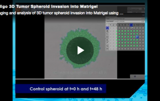
Video: 3D tumor spheroid invasion into Matrigel using u-bottom 96-well ULA plates. Examine drug effects on tumor spheroids as a model system for tumor invasion.
Video: 3D tumor spheroid invasion into Matrigel using u-bottom 96-well ULA plates. Examine drug effects on tumor spheroids as a model system for tumor invasion.
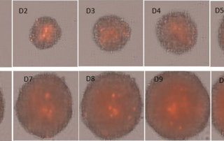
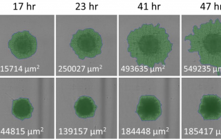
Kinetically monitor multicellular tumor spheroid (MCTS) growth & Propidium Iodide (PI) dead cell signal on U87MG Glioblastoma over mulitple days
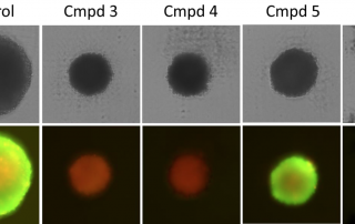
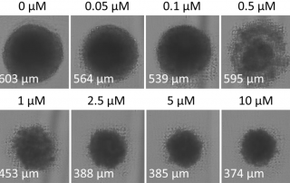
Measure the effects of a panel of drugs on the viability of U87MG Glioblastoma MCTS using calcein AM and Propidium Iodide fluorescent staining
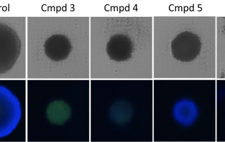
Monitor the effects of a panel of drugs on the apoptosis of U87MG Glioblastoma MCTS using Caspase 3/7 and Hoechst fluorescent staining
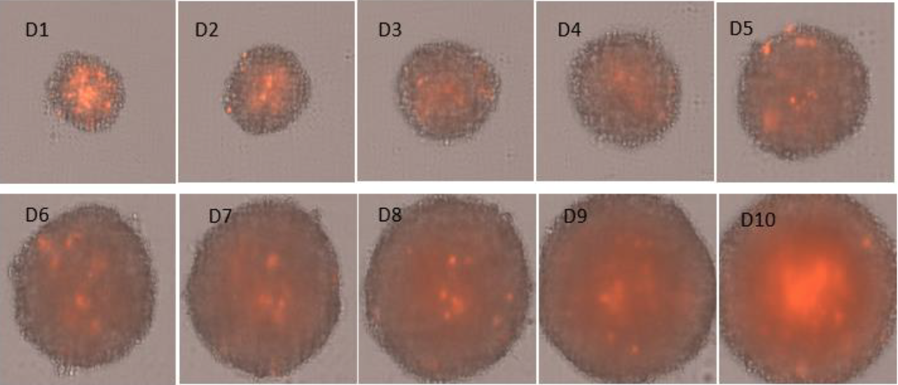
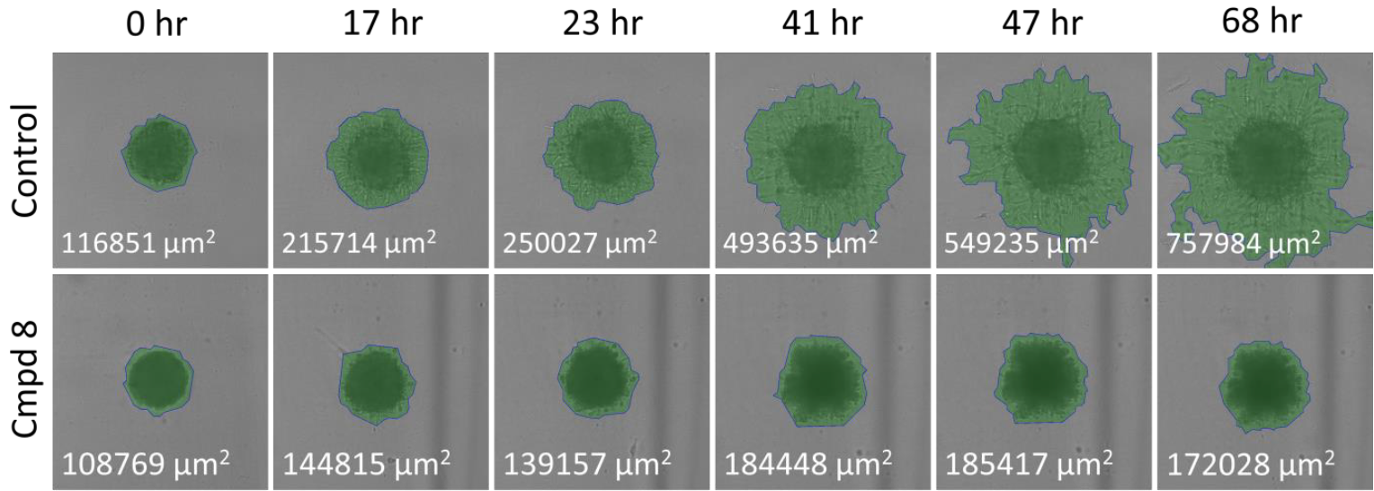
Monitor the effects of a panel of drugs on the invasion of U87MG Glioblastoma MCTS into Basement Membrane Extract (BME) Matrigel
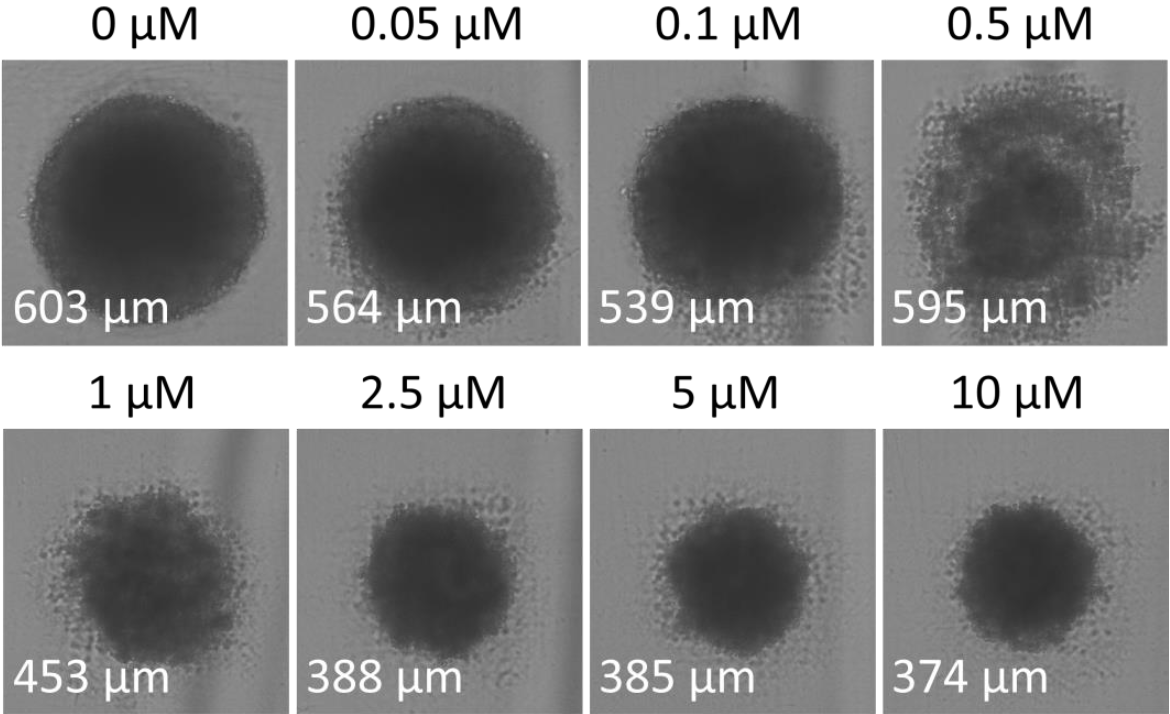
Using the bright field imaging, the Celigo will rapidly provide multicellular tumor spheroid images and diameters of treated U87MG MCTS
Using the bright field imaging, the Celigo will rapidly provide multicellular tumorspheroid images and diameters of treated MCTS in a 96-well plate
Join us on Thursday, June 7th for an informational webinar by Somaieh Hedayat of The Institute of Cancer Research. She will discuss different approaches to recapitulate the metastatic niche and compare PDOs drug responses observed in the laboratory ex vivo with clinical responses observed in patients and how those techniques will allow the study of PDO drug responses in the pre-clinical setting strengthening their value as a platform for drug screening, discovery and development. About Somaieh Hedayat: Somaieh Hedayat is working towards a PhD on how the tissue microenvironment can affect the response of metastatic colorectal cancer cells to antiangiogenic therapies. Prior [...]
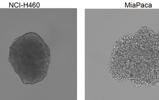
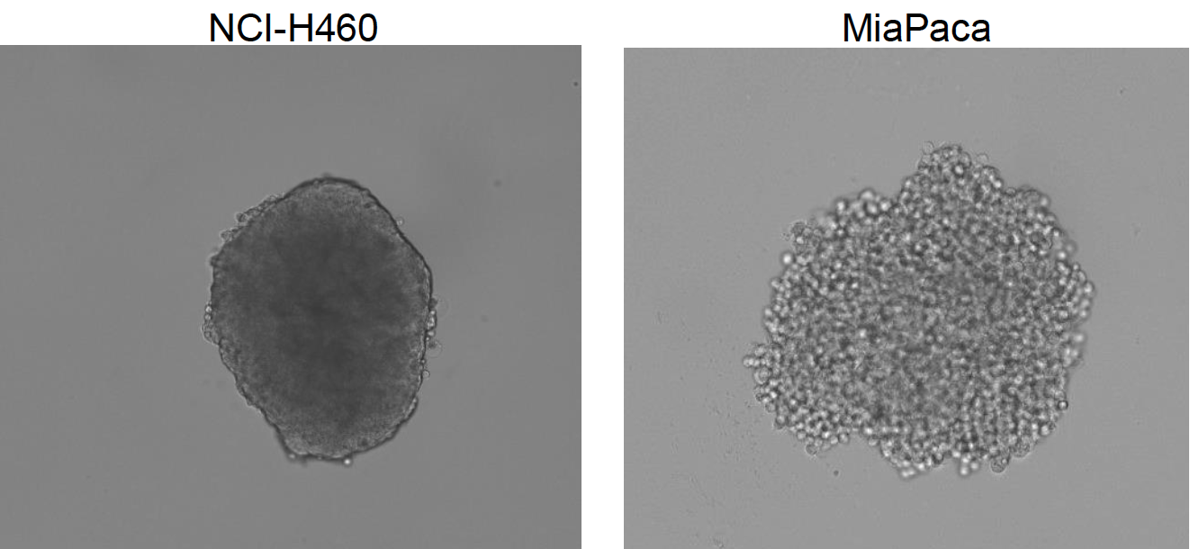
Recent publications have suggested that using 3D tumor spheroids is a more predictive model for preclinical research. Nexcelom Bioscience has developed a standardized microplate method for rapid generation, imaging and analysis of 3D tumor spheroids using the Celigo image cytometer. 40 cancer cell lines' ability to form spheroids, optimal seeding densities and culture conditions have been established. Protocols measuring tumor growth, viability, migration and invasion have been utilized by many researchers for routine preclinical drug studies. If you are interested to learn more about how your research might benefit from working with 3D tumor spheroids, this is a great place to start! [...]
It's White Paper Wednesday! Read our featured white paper: 3D Tumor Spheroid Analysis Method for HTS Drug Discovery using Celigo Imaging Cytometer U87MG cells were used to create tumorspheres in 384-well plates that were subsequently analyzed by imaging. The data illustrate that reproducible 3D spheroids can be formed in 384-well plates. Fluorescent viability studies were carried out with the imager using pixel intensity analysis. Moreover, the assay was validated for drug screen using various drug compounds that have shown anti-proliferative effects. Together, these data demonstrate that the tumorsphere formation assay can be developed, validated and used for high-throughput anti-cancer compound [...]
