Introduction
Propofol, a widely used general anesthetic, has been shown to produce neurotoxicity in neonatal animal experiments [1-5]. The possible detrimental effects of anesthetics, particularly in developing brains, require further study, as recent studies suggest that general anesthesia given to children younger than four years of age can increase the chances of learning disabilities later in life [6]. Here, researchers used embryonic neural stem cells to investigate the neurotoxic effects of propofol in vitro.
Materials and Methods
Cell culture and drug exposure
Embryonic neural stem cells were collected from embryonic day 16 Sprague-Dawley rats. Resulting cells were cultured in 96-well plates in DMEM/F12 medium plus N2. On Day 8 in vitro, the cells were exposed to various concentrations (0-600 μM) propofol for 24 hours.
Apoptosis/necrosis analysis
Cells were washed and stained with a mixture of FITC-labeled Annexin V and propidium iodide (PI). Cellometer Vision was used to image and analyze all cultures. Cells positive for Annexin V staining were considered apoptotic, cells positive for PI staining were considered necrotic, and cells stained with neither were considered live.
*Note: For more detailed Materials and Methods and a complete account of the entire study, please refer to the original manuscript [http://www.ncbi.nlm.nih.gov/pubmed/24704589]
Results
- 50 μM propofol given for 24 hours showed an increase in percentage of apoptotic cells and a decrease in percentage of live cells as compared to control.
- There was no significant change in percentage of necrotic cells across the conditions.
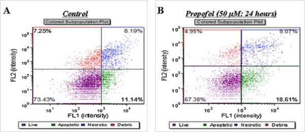
Figure 1. Effects of propofol on neural stem cell health. Scatter plots demonstrate the percentage of live cells (purple), apoptotic cells (Annexin V positive; green), and necrotic cells (PI positive; blue) for control (A) and 50 μM propofol treated (B) cultures.
Conclusions
- Clinically-relevant doses of propofol given long-term induce toxicity in embryonic neural stem cells in vitro through mechanisms of oxidative stress and mitochondrial dysfunction.
- Because anesthesia for children is typically extrapolated from adult doses, it is important to more fully understand the unique vulnerabilities to anesthesia children may possess.
- Using embryonic neural stem cells is a valuable model to better understand these differences and help prevent anesthesia-induced neurotoxicity in pediatric populations.
References
- Vutskits, L., et al., Clinically relevant concentrations of propofol but not midazolam alter in vitro dendritic development of isolated gamma-aminobutyric acid-positive interneurons. Anesthesiology, 2005. 102(5): p. 970-6.
- Cattano, D., et al., Subanesthetic doses of propofol induce neuroapoptosis in the infant mouse brain. Anesth Analg, 2008. 106(6): p. 1712-4.
- Pesic, V., et al., Potential mechanism of cell death in the developing rat brain induced by propofol anesthesia. Int J Dev Neurosci, 2009. 27(3): p. 279-87.
- Bercker, S., et al., Neurodegeneration in newborn rats following propofol and sevoflurane anesthesia. Neurotox Res, 2009. 16(2): p. 140-7.
- Milanovic, D., et al., Regional and temporal profiles of calpain and caspase-3 activities in postnatal rat brain following repeated propofol administration. Dev Neurosci, 2010. 32(4): p. 288-301.
- Wilder, R.T., et al., Early exposure to anesthesia and learning disabilities in a population-based birth cohort. Anesthesiology, 2009. 110(4): p. 796-804.


Leave A Comment