| Purpose | Perform apoptosis assay on MDA-MB-231 and Jurkat cells |
| Existing Method(s) | Flow cytometry |
| Target Cell Type | MDA-MB-231 and Jurkat cells |
| Experiment Plan | Scan plate using Green, Bright field and Blue channels |
| Hypothesis | By measuring the number of Caspase 3/7 positive cells, we can determine the percent of apoptotic cells in the population |
Celigo Setup
| Plate Type | Greiner 655090 96-well black wall clear bottom |
| Scan Channels | Green, Brightfield and Blue |
| Resolution | 1 µm/pixel |
| Scan Area | Whole well |
| Analysis Method | Target 1 + 2 + Mask |
| Scan Frequency | End Point |
| Scan Time | ~15 minutes |
Assay Protocol and Plate Setup
Goal:
Detect and quantify apoptotic cells using Caspase 3/7 and Hoechst staining in adherent MDA-MB-231 and suspension Jurkat cell lines
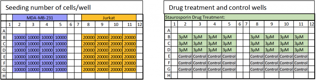
Protocol:
- Seed MDA-MB-231 at 10,000 cells/well and Jurkat cells at 20,000 cells/well and allow to incubate overnight
- Add Staurosporine at 3 µM final concentration per well and allow to incubate for 4-6 hours
- After incubation is completed, prepare in PBS a 2X concentration of Caspase 3/7 and Hoechst
- Nexcelom, Cat# CSK-V0003-1
- Remove 100 µL of media from all plate wells.
- Add 100 µL of 2X concentration of Caspase 3/7 and Hoechst and incubate for 30 mins at 37° C
- Image the plate using the Celigo image cytometer
Results
Drug-treated MDA-MB-231 and Jurkat cells showed an increase in Caspase 3/7 positive cells
- Total number of nucleated cells was determined by counterstaining the cells with Hoechst
- Total number of apoptotic cells was determined by counting the nucleated cells stained with green Caspase 3/7 reagent
- Determined the percent of apoptotic-positive cells
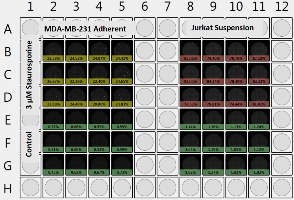
Plate-Level Data View allows a quick observation of the total number of cells and green Caspase 3/7 positive cells, as well as percent Caspase 3/7 positive cells. Currently displaying percent Caspase 3/7.
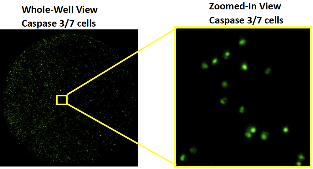
Whole-well view allows high-resolution observation of images and at zoomed levels.
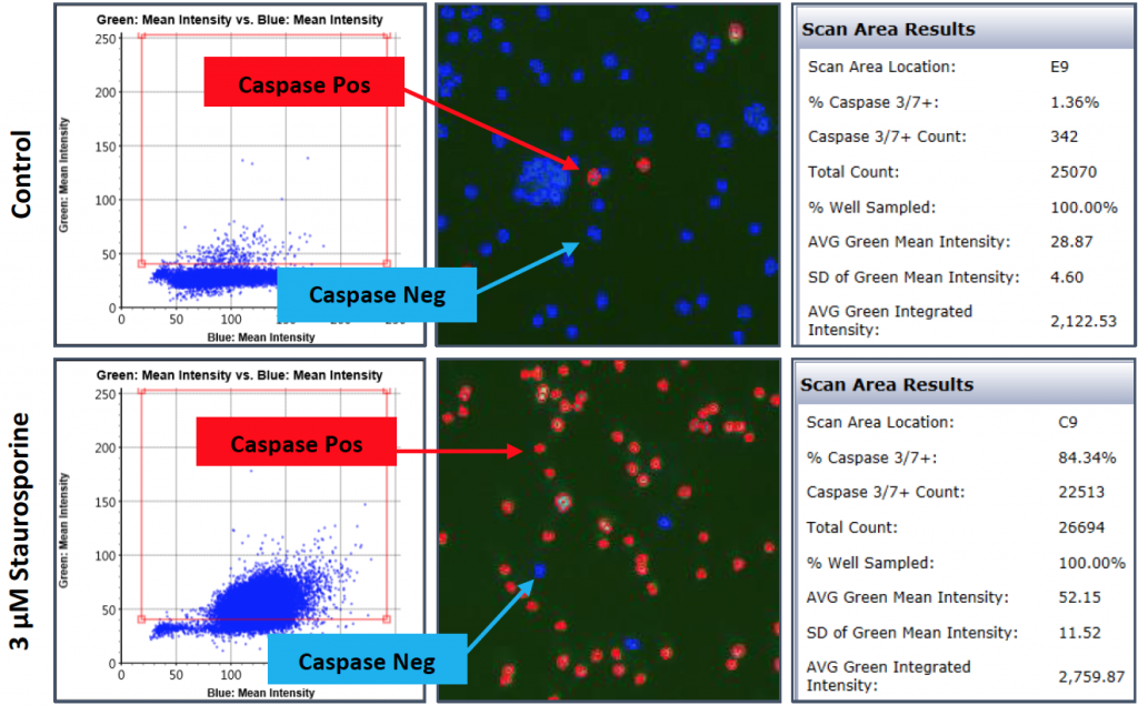
Gate Plots for Caspase 3/7 Positive Cells:
Apoptosis Caspase 3/7 gating analysis of an MDA-MB-231 and Jurkat cells
MDA-MB-231 Adherent:
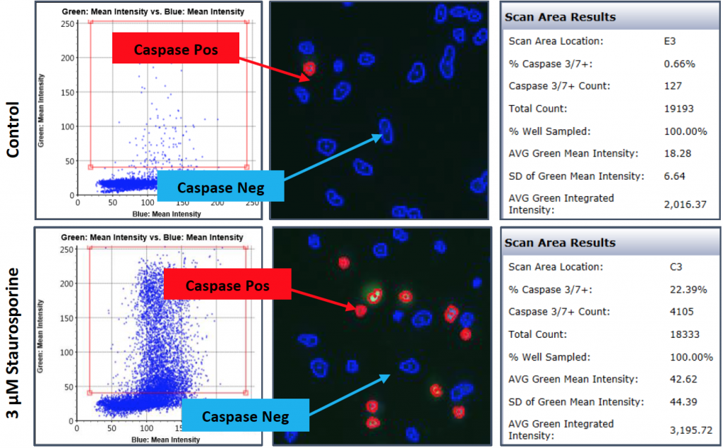
Jurkat Suspension:
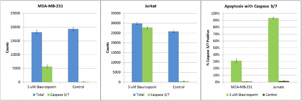
Graphs
- Cell counts for adherent, MDA-MBA-231 and suspension, Jurkat cells plotted
- Using Hoechst cell counts as a total, percent Caspase 3/7 positive cells are plotted on bar graphs
Conclusion
- The Celigo successfully performed Caspase 3/7 apoptosis assay using MDA-MB-231 and Jurkat cell lines
- Acquisition of high-resolution bright field, Caspase 3/7, and Hoechst fluorescent images of an entire 96 well plate took ~ 15 minutes
- Performing an endpoint apoptosis assay using Caspase 3/7 with Hoechst allows for the enumeration of the total number of nucleated cells and the total number of Caspase 3/7 positive cells, as well as to determine the percent of apoptosis.