Transcript: Image-based Analysis of Apoptosis with Annexin V-FITC / PI
Cellometer Vision CBA is a simple, image-based instrument optimized for analysis of cell-based assays for apoptosis.
For this apoptosis experiment, Jurkat cells are incubated for 15 minutes with Annexin V-FITC and propidium iodide. To analyze the cells, pipette 20µl of sample into the Cellometer counting chamber. Insert the chamber into the Vision CBA instrument.
Select the Apoptosis Assay from the drop-down menu. Enter the Sample ID in the appropriate field, then click count.
In less than 3 minutes, the Vision CBA takes multiple cell images and analyzes the images based on pre-set parameters for the Apoptosis Assay selected.
When cell imaging and counting is complete, the initial results table displays total cells counted, cell concentration, and mean cell diameter.
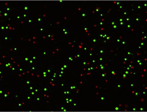
The brightfield counted image can be viewed for confirmation that cellular debris has not been counted. Fluorescent images show Annexin V-FITC positive apoptotic cells and the PI positive necrotic cells.
Click export. After defining a location for the export file, an optimized data report will be displayed. Apoptosis data is displayed as a standard scatter plot and also a colorized plot based on cell population. The automatic gating can be manually adjusted. Associated data tables update automatically to reflect revised percentages.
Users can choose to print a second report containing both brightfield and fluorescent images. Data tables and cell images can be easily exported for use in presentations and publications.
After removing the disposable Cellometer cell-counting chamber, the Vision CBA is ready to analyze the next sample. No washing or instrument set-up is required.
Cellometer Vision CBA offers researchers a simple, image-based alternative for a growing number of cell-based assays. Experienced applications specialists are available to demonstrate the Vision CBA in your laboratory. Visit www.nexcelom.com or contact Nexcelom today to learn more.


Leave A Comment