Comparison of Trypan Blue and Fluorescence-Based Viability Detection Methods Via Morphological Observation

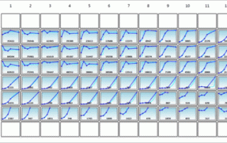
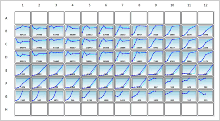
It's White Paper Wednesday! Read our featured white paper: Comparison of Trypan Blue and Fluorescence-Based Viability Detection Methods Via Morphological Observation Determining cell viability is a vital component in many biological experiments that range from standard cell culture to the investigation of pharmacological agents on tumor cells. One of the earliest and most common methods for measuring cell viability is the trypan blue (TB) exclusion assay [1, 2]. Over the last two decades, there have been various publications on comparing TB exclusion and fluorescence-based cellular viability assays [1, 3-5]. Previous results have shown that in a time-course measurement, TB exclusion [...]

Automated Method for Determination of Infectious Dose (TCID50) using Celigo Imaging Cytometer

It's White Paper Wednesday! Read our featured white paper: Automated Method for Determination of Infectious Dose (TCID50) using Celigo Imaging Cytometer Nexcelom's Celigo imaging cytometer has been applied to provide automated, rapid assessment of viral infectivity in a range of plate formats [4]. Using f-theta optics, Celigo provides high quality, whole-well images using bright field and/or fluorescent illumination. Automated segmentation and analysis provide quantitative and objective output of CPE based on characteristic changes to the host cell monolayer. Download our white paper »

Characterization of Breast Cancer Drugs via Mammosphere Morphometric Analysis Using Celigo Imaging Cytometer

It's White Paper Wednesday! Read our featured white paper: Characterization of Breast Cancer Drugs via Mammosphere Morphometric Analysis Using Celigo Imaging Cytometer A panel of cytotoxic drugs, including doxorubicin and paclitaxel were used to study their effects on various breast cancer cell lines such as MDA-MD-436, MCF-7, SKBR3 and MDA-MB-231. Results show that the Colony Counting application can also be used to evaluate the clonogenicity and self-renewal of cancer stem/tumor-initiating cells by automatically analyzing mammosphere populations. The Colony Counting application on Celigo provides an efficient, reproducible and automated method for assessing the number, size, and morphology of cancer spheroids within [...]

Measuring Physiological and Metabolic Characteristics of Yeast Health for Beer Fermentation Samples using the Cellometer Vision

It's White Paper Wednesday! Read our featured white paper: Measuring Physiological and Metabolic Characteristics of Yeast Health for Beer Fermentation Samples using the Cellometer Vision Characteristics such as the viability, vitality, glycogen, neutral lipid, and trehalose content of yeast samples must be measured to better understand changes in physiological and metabolic status during fermentation [4-9]. Monitoring changes of these parameters during fermentation can improve current production processes, and a subset of these parameters (glycogen, neutral lipid, and trehalose content) has been shown to play an important role in predicting yeast viability both during and after fermentation [10, 11]. Download our [...]

By |2021-06-15T19:52:03+00:00April 8th, 2015|Categories: Cellometer, Cellometer Application News, Instrument|Tags: , , , , |0 Comments

Comparing Fluorescence-Based Viability Detection Method using the Cellometer Vision

It's White Paper Wednesday! Read our featured white paper: Comparing Fluorescence-Based Viability Detection Method using the Cellometer Vision In this work, Cellometer Vision was employed to demonstrate rapid fluorescence-based viability measurements and the comparison of various fluorescent staining methods. First, fluorescent nucleic acid stains that examine membrane integrity were tested and validated by comparing against the standard trypan blue exclusion method. Similarly, fluorescent enzymatic stains that examine metabolic activities were tested and validated against the standard trypan blue exclusion method. Third, nucleic acid and enzymatic stains were compared by measuring viabilities of Jurkat cells incubated at different temperatures of water [...]

Bright field and Fluorescent Image Analysis for Screening Applications using Celigo

Generate growth curves over time and monitor cell counts and confluence at the individual well level.

Detection of Multiplexed GFP Reporters in Primary Articular Chondrocyte Cultures Using Cellometer Vision Image Cytometer

It's White Paper Wednesday! Read our featured white paper: Detection of Multiplexed GFP Reporters in Primary Articular Chondrocyte Cultures Using Cellometer Vision Image Cytometer In this work, we have developed an image cytometry method for detecting and monitoring the cell expansion and differentiation of articular chondrocytes in primary culture. First, the feasibility of utilizing image cytometry for detection of fluorescent is shown by comparing measured fluorescent positive cell populations to flow cytometry. Next, articular chondrocyte cultures were established in multi-well plates from either single or Cyan/eGFP double reporter mouse lines and grown for 20 days to test the utility of the [...]

By |2021-06-15T20:34:28+00:00February 25th, 2015|Categories: Cellometer, Cellometer Application News, Instrument|Tags: , , , , |0 Comments

The Historical Development of the Hemacytometer

It's White Paper Wednesday! Read our featured white paper:The Historical Development of the Hemacytometer The hemacytometer has been an essential tool for hematologists, medical practitioners, and biologists for over a century. Depending on where it is being used, the word has multiple spellings such as hemacytometer, hemocytometer, haemacytometer, or haemocytometer, but for consistency purposes the word “hemacytometer” will be used in this review. The prefix “hema”, “hemo”, “haema”, or “haemo” means blood, while “cytometer” meant a device to measure cells. The device was initially used by medical practitioners to analyze patient blood samples, which was the initial spark that created [...]

Accurately Count PBMC and Measure Viability in Presence of Residual RBC

It’s White Paper Wednesday! This month’s featured white paper: Accurately Count PBMC and Measure Viability in Presence of Residual RBC In this work, we have developed an image cytometry method for detecting and monitoring the cell expansion and differentiation of articular chondrocytes in primary culture. First, the feasibility of utilizing image cytometry for detection of fluorescent is shown by comparing measured fluorescent positive cell populations to flow cytometry. Next, articular chondrocyte cultures were established in multi-well plates from either single or Cyan/eGFP double reporter mouse lines and grown for 20 days to test the utility of the fluorescence-based image cytometry system. [...]

Go to Top