Video: Celigo Multi-Channel Imaging Cell Cytometer
Celigo is a multi-channel imaging cell cytometer designed for scientists performing bench-top experiments in micro-well plates.
Because the Celigo can image suspension and adherent cells, there is no need to trypsinize adherent cells or colonies for flow-based analysis.
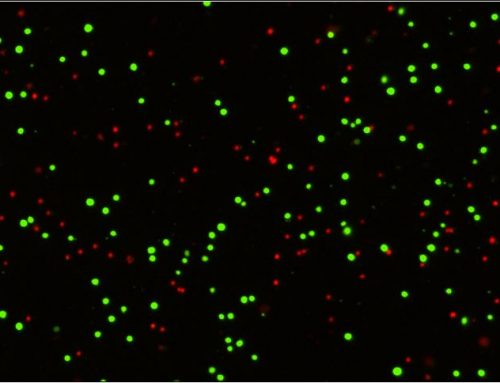
We are able to obtain population and single cell data by imaging every cell in every well in both bright field and fluorescent channels.
The proprietary technology in the system makes it possible to obtain high quality images at high throughput.
The system is run by software that is designed with a simple work flow to be easily used by everyday bench biologists.
Since 2010, the Celigo has been effectively used by scientists in academia and industry to study and perform drug screening investigations utilizing hundreds of cells lines and thousands of compounds.
By using the direct bright field cell counting method, they are able to perform cell cytotoxicity, cell survival, and proliferation assays on adherent cell lines in multi-well plates. Downstream fluorescent functional assays such as cell cycle, apoptosis, and viability are also routinely performed to examine drug effects on cell health.
Over the last few years Celigo has also been effectively used to monitor iPSC reprogramming, stem cell pluripotency and differentiation. Quantifying the transduction efficiencies, looking at the expression of Yamanaka factors and monitoring the expression of pluripotency markers, are just some of the assays that are routinely performed on the Celigo while monitoring iPSC reprogramming. The Celigo can track the evolution, number, and growth of iPSC colonies in a single plate throughout the entire reprogramming process.
For tumorspheres, the Celgo has recently been effectively utilized for target validation and evolution of therapeutics in 3D tumor cultures. In addition to imaging and examining the inhibition or growth of tumor spheroids, the instrument is capable of examining the cell invasion into matrigel from the spheroid. The Celigo’s ability to image colonies and spheroids in flat bottom and u-bottom plates makes it a uniquely versatile instrument.
-These and many other studies have been published in high-impact peer-reviewed journals such as Nature, Breast Cancer Research, BMC Biology, and others.
If you are interested in learning more, please contact us to speak with one of our Field Applications Scientists, who will be happy to answer any questions you may have about the Celigo S Image Cytometer and how it can benefit your research. Our Applications Scientists can also arrange a free on-site technical seminar or demonstration for you and your colleagues.


Leave A Comment