Transcript: How to Quickly and Easily Measure Caspase-8 Activity
Because Caspase-8 is activated upon initiation of the extrinsic apoptosis pathway, measurement of active Caspase-8 is of particular interest.
In this experiment, 1µl of a FITC-conjugated Caspase-8 inhibitor (that specifically binds active Caspase-8 within the cell) was incubated with 300 µl of cell sample for 1 hour then analyzed with the Cellometer Vision CBA Analysis System.
To analyze a sample for active Caspase-8, select the Caspase Assay from the drop-down menu, enter the Sample ID, then click Count. Cell images are captured and analyzed based on preset parameters for the Caspase-8 assay.
A report showing the total cell count, concentration, and mean diameter is automatically displayed.
Clicking Export brings up the Caspase positive / Caspase negative cell population histogram.
The associated data table displays the percent and concentration of cells for each population.
Cell images can be viewed to check cell morphology and review counted cells. In the bright field counted image, all counted cells are outlined in green.
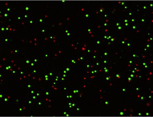
The fluorescent image indicates cells positive for active Caspase-8.
The diameter of each cell is measured to generate a size distribution histogram.
Control gates can be applied to treated samples to measure increases in the active Caspase-8 population
Visit www.nexcelom.com for more information on the Caspase-8 Assay Kit, other cell-based assay kits, and the Cellometer Vision CBA Analysis System.


Leave A Comment