Celigo Experiment Examples
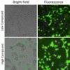
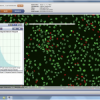
Celigo Experiment Examples are 2D and 3D assays that have been successfully performed on the Celigo Image Cytometer. This bench-top, bright field and fluorescent imaging system provides high speed, fully automated imaging and quantification of adherent and suspension cells in any format (6, 12, 24, 48, 96, 384, 1536-well plates, T flasks, and slides).
Celigo’s versatility allows for a wide range of assays that can be routinely and efficiently carried out during everyday lab operations. From cell line development to functional assays to 3D tumor spheroid analysis, there is no doubt that Celigo can help you take your experiments to the next level. Read or download any of the assays below to better understand how Celigo can be used in your lab.
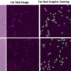
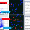
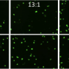
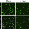
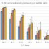
Immuno-Oncology
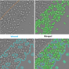
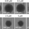
Migration/Invasion
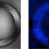
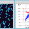
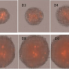
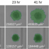
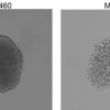
3D Models
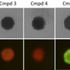
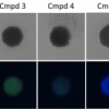
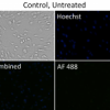
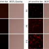
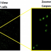
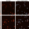
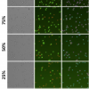
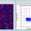
Fluorescent Assays