A Novel Imaging Cytometry Method for Immunophenotyping
Leo L. Chan, Xuemei Zhong, Jean Qiu, Peter Y. Li, Bo Lin
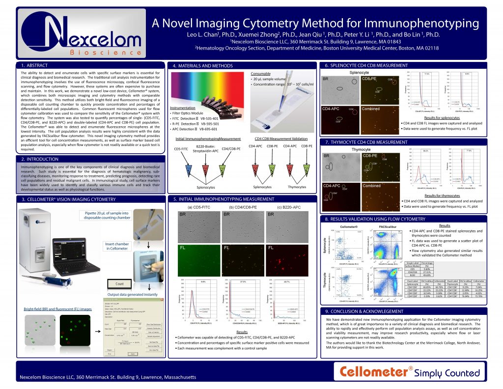
The ability to detect and enumerate cells with specific surface markers is essential for clinical diagnosis and biomedical research. The traditional cell analysis instrumentation for immunophenotyping involves the use of fluorescence microscopy, confocal fluorescence scanning, and flow cytometry. However, these systems are often expensive to purchase
and maintain. In this work, we demonstrate a novel low-cost device, Cellometer® system, which combines both microscopic imaging and cytometry methods with comparable detection sensitivity. This method utilizes both bright-field and fluorescence imaging of a disposable cell counting chamber to quickly provide concentration and percentages of
differentially-labeled cell populations. Common fluorescent microspheres used for flow cytometer calibration was used to compare the sensitivity of the Cellometer® system with flow cytometry. The system was also tested to quantify percentages of single- (CD5-FITC, CD4/CD8-PE, and B220-APC) and double-labeled (CD4-APC and CD8-PE) cell population.
The Cellometer® was able to detect and enumerate fluorescence microspheres at the lowest intensity. The cell population analysis results were highly consistent with the data generated by FACScalibur flow cytometer. This novel imaging cytometry method provides an efficient tool for cell concentration measurements, as well as surface marker based cell
population analysis, especially when flow cytometer is not readily available or a quick test is required.