Apoptosis Analysis of Jurkat Cells using the Cellometer Vision
Leo L. Chan, Tim Smith, Alnoor Pirani, Emily Lyettefi, Ning Lai, Jean Qiu, and Bo Lin
We demonstrate a rapid and cost-effective method for apoptosis analysis at various stages of Jurkat cells using the Cellometer® Vision. This method has the ability to eliminate many known issues caused by manual hemacytometer and flow cytometer. By using Cellometer® Vision, the assay time for obtaining apoptosis result is greatly reduced, which is significant for research development in academia and industry.
The current methods for apoptosis analysis utilizes standard flow cytometry, but several inherent issues are attached. Standard flow cytometry is expensive, large in size, and require considerable amount of maintenance. In addition, most of the flow cytometers do not have imaging capabilities, which often generate some uncertainties in the fluorescence results obtained. Recently, a novel imaging cytometry platform, equivalent of combining
fluorescent microscope and flow cytometry, has been developed by Nexcelom Bioscience (Lawrence, MA). This new device allows rapid analysis using inexpensive disposable counting chambers that require only 20 μl of samples. This system allows automated cell image acquisition and processing with a novel counting algorithm for accurate and consistent measurement of cell population and viability on a variety of cell types
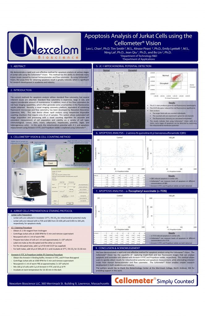
(immunological, cancer, stem, insect, adipocytes, hepatocytes, platelets, algae, and heterogeneous cells). In this work, the mitochondrial potential with JC-1 and apoptosis measurement using Annexin V-FITC and Propidium Iodide is explored using Cellometer®.