Comparison of label-free cell cytotoxicity image cytometric detection method to CellTiter-Glo®
Roopal Patel, Olivier Déry, Leo Li-Ying Chan, and Gina Wei
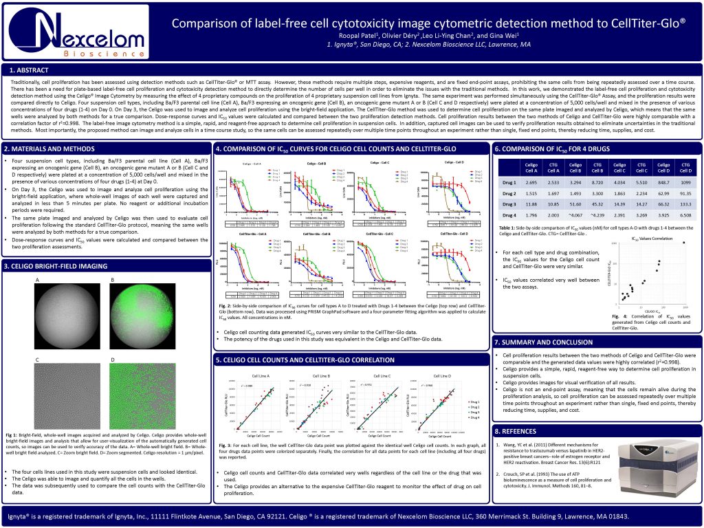
Traditionally, cell proliferation has been assessed using detection methods such as CellTiter-Glo® or MTT assay. However, these methods require multiple steps, expensive reagents, and are fixed end-point assays, prohibiting the same cells from being repeatedly assessed over a time course.
There has been a need for plate-based label-free cell proliferation and cytotoxicity detection method to directly determine the number of cells per well in order to eliminate the issues with the traditional methods. In this work, we demonstrated the label-free cell proliferation and cytotoxicity
detection method using the Celigo® Image Cytometry by measuring the effect of 4 proprietary compounds on the proliferation of 4 proprietary suspension cell lines from Ignyta. The same experiment was performed simultaneously using the CellTiter-Glo® Assay, and the proliferation results were compared directly to Celigo. Four suspension cell types, including Ba/F3 parental cell line (Cell A), Ba/F3 expressing an oncogenic gene (Cell B), an oncogenic gene mutant A or B (Cell C and D respectively) were plated at a concentration of 5,000 cells/well and mixed in the presence of various
concentrations of four drugs (1-4) on Day 0. On Day 3, the Celigo was used to image and analyze cell proliferation using the bright-field application. The CellTiter-Glo method was used to determine cell proliferation on the same plate imaged and analyzed by Celigo, which means that the same
wells were analyzed by both methods for a true comparison. Dose-response curves and IC50 values were calculated and compared between the two proliferation detection methods. Cell proliferation results between the two methods of Celigo and CellTiter-Glo were highly comparable with a
correlation factor of r 2=0.998. The label-free image cytometry method is a simple, rapid, and reagent-free approach to determine cell proliferation in suspension cells. In addition, captured cell images can be used to verify proliferation results obtained to eliminate uncertainties in the traditional
methods. Most importantly, the proposed method can image and analyze cells in a time course study, so the same cells can be assessed repeatedly over multiple time points throughout an experiment rather than single, fixed end points, thereby reducing time, supplies, and cost.