A high-throughput image cytometry-based screening method for the detection of IL2-induced peripheral blood mononuclear cell-mediated cytotoxicity
Leo L. Chan, Kelsey J, McCulley, Pinaki P. Banerjee
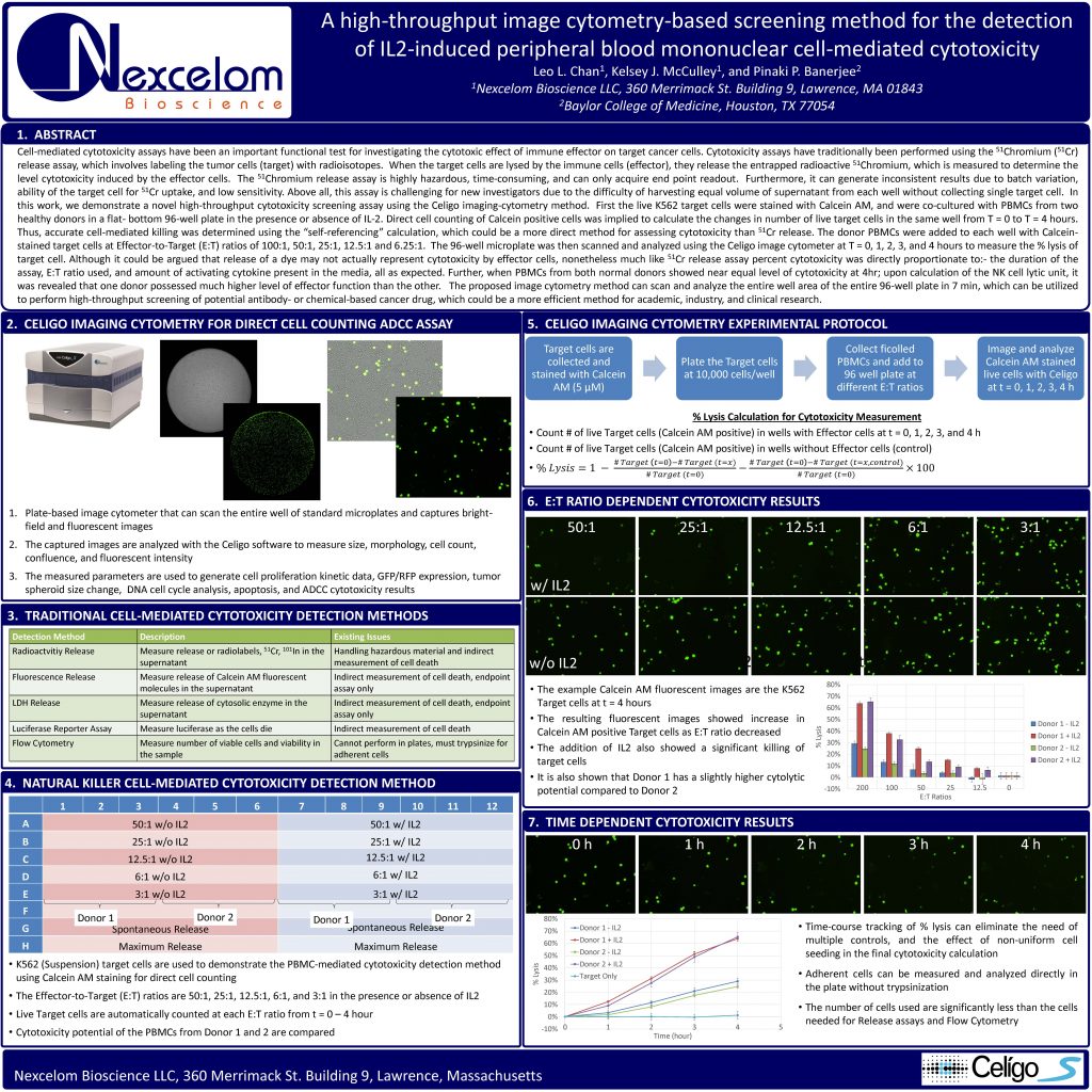
Cell-mediated cytotoxicity assays have been an important functional test for investigating the cytotoxic effect of immune effector on target cancer cells. Cytotoxicity assays have traditionally been performed using the
51Chromium (51Cr) release assay, which involves labeling the tumor cells (target) with radioisotopes. When the target cells are lysed by the immune cells (effector), they release the entrapped radioactive 51Chromium, which is measured to determine the level cytotoxicity induced by the effector cells. The 51Chromium release assay is highly hazardous, time-consuming, and can only acquire end point readout. Furthermore, it can generate inconsistent results due to batch variation, ability of the target cell for 51Cr uptake, and low sensitivity. Above all, this assay is challenging for new investigators due to the difficulty of harvesting equal volume of supernatant from each well without collecting single target cell. In this work, we demonstrate a novel high-throughput cytotoxicity screening assay using the Celigo imaging-cytometry method. First the live K562 target cells were stained with Calcein AM, and were co-cultured with PBMCs from two healthy donors in a flat- bottom 96-well plate in the presence or absence of IL-2. Direct cell counting of Calcein positive cells was implied to calculate the changes in number of live target cells in the same well from T = 0 to T = 4 hours.
Thus, accurate cell-mediated killing was determined using the “self-referencing” calculation, which could be a more direct method for assessing cytotoxicity than 51Cr release. The donor PBMCs were added to each well with Calceinstained target cells at Effector-to-Target (E:T) ratios of 100:1, 50:1, 25:1, 12.5:1 and 6.25:1. The 96-well microplate was then scanned and analyzed using the Celigo image cytometer at T = 0, 1, 2, 3, and 4 hours to measure the % lysis of target cell. Although it could be argued that release of a dye may not actually represent cytotoxicity by effector cells, nonetheless much like 51Cr release assay percent cytotoxicity was directly proportionate to:- the duration of the assay, E:T ratio used, and amount of activating cytokine present in the media, all as expected. Further, when PBMCs from both normal donors showed near equal level of cytotoxicity at 4hr; upon calculation of the NK cell lytic unit, it was revealed that one donor possessed much higher level of effector function than the other. The proposed image cytometry method can scan and analyze the entire well area of the entire 96-well plate in 7 min, which can be utilized to perform high-throughput screening of potential antibody- or chemical-based cancer drug, which could be a more efficient method for academic, industry, and clinical research.