A Novel image based cytometry analysis for measuring cell migration in wound healing assay
Leo L. Chan, Scott Cribbes, Sarah Kessel, Olivier Dery, Dmitry Kuksin, and Jean Qiu
Cell migration is a multi-stepped, highly complex process that is involved in normal processes of cell proliferation and homeostasis, but also is exaggerated in the pathologies of metastasis and tumour invasion. The
coordination of events has been studied at the molecular, biochemical and biophysical level for nearly 50 years. One assay which has been used throughout is the wound-healing or scratch assay. Simply defined, a
monolayer of cells are grown and a border is introduced either by scratching through this monolayer to create a wound or by removing a physical barrier. The movement of cells over the margin and into the newly
created space is measured. Additional information that can be gleaned from this type of assay may also be cell morphology and polarity. This biological process can substantially differ depending on the origin of the cells,
the matrix they are grown on, the composition of the media and any compounds/nucleic acids that may be added as part of a screen, therefore it is important to set up a robust assay that will allow for many
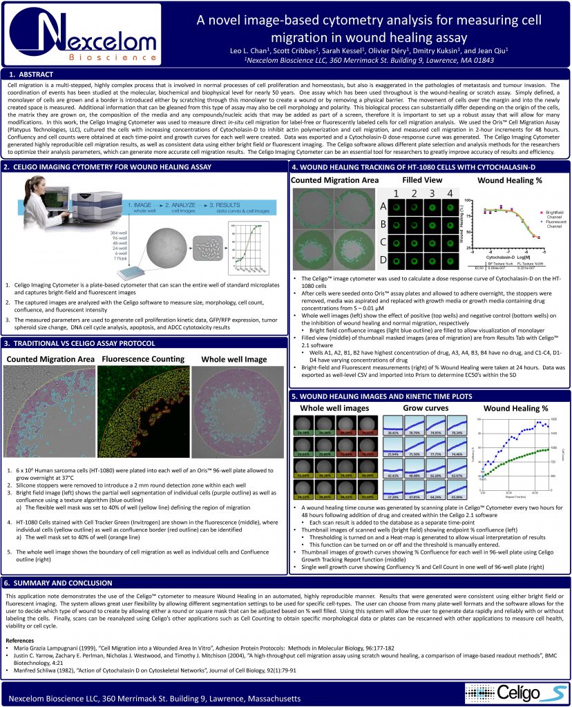
modifications. In this work, the Celigo Imaging Cytometer was used to measure direct in-situ cell migration for label-free or fluorescently labeled cells for cell migration analysis. We used the Oris™ Cell Migration Assay
(Platypus Technologies, LLC), cultured the cells with increasing concentrations of Cytocholasin-D to inhibit actin polymerization and cell migration, and measured cell migration in 2-hour increments for 48 hours.
Confluency and cell counts were obtained at each time-point and growth curves for each well were created. Data was exported and a Cytocholasin-D dose-response curve was generated. The Celigo Imaging Cytometer
generated highly reproducible cell migration results, as well as consistent data using either bright field or fluorescent imaging. The Celigo software allows different plate selection and analysis methods for the researchers
to optimize their analysis parameters, which can generate more accurate cell migration results. The Celigo Imaging Cytometer can be an essential tool for researchers to greatly improve accuracy of results and efficiency.