A Rapid Image Cytometry Method for Quantification of Canine Stromal Vascular Fraction Cells
Leo L. Chan, Donald A. Cohen, Dmitry Kuksin, Benjamin D. Paradis, and Jean Qiu
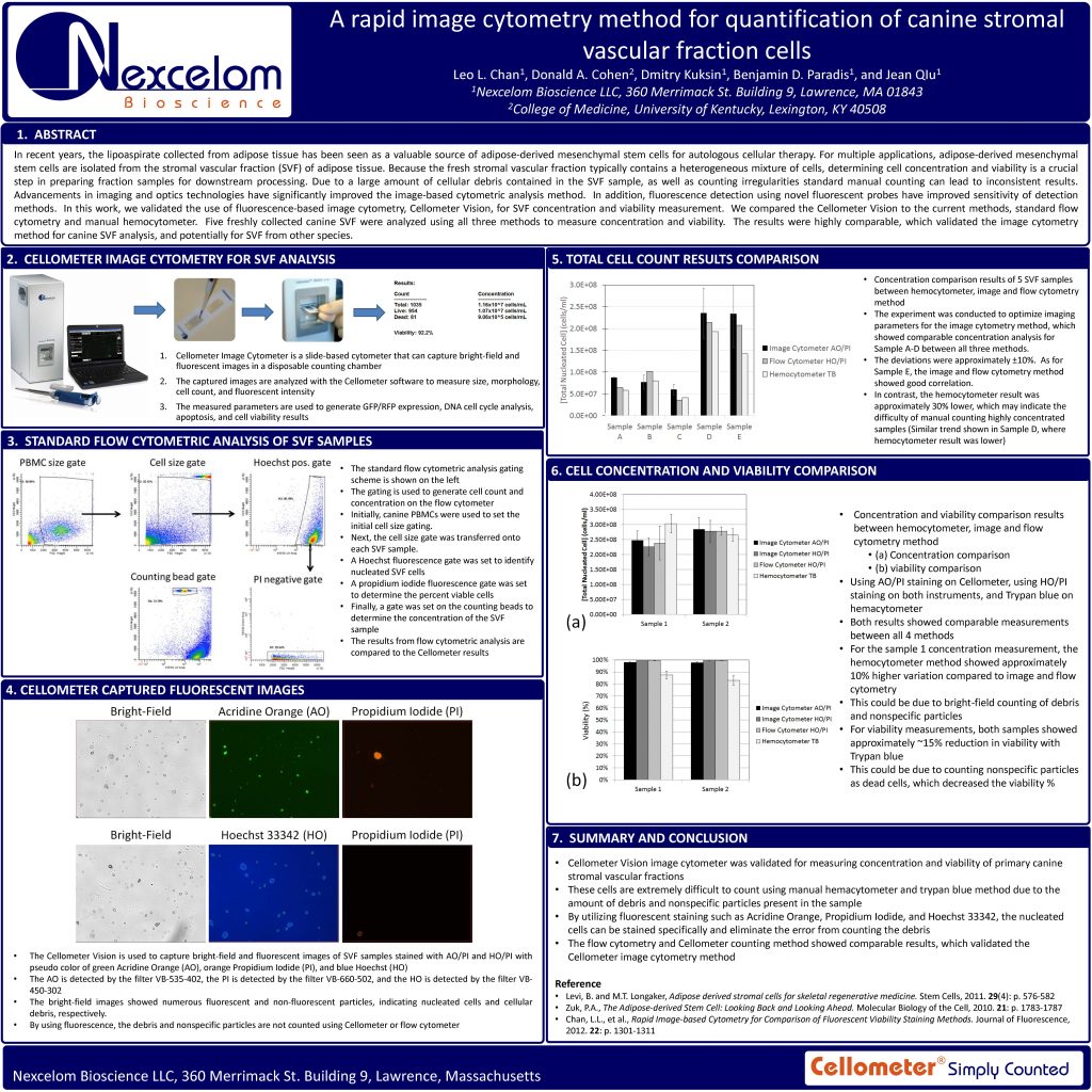
In recent years, the lipoaspirate collected from adipose tissue has been seen as a valuable source of adipose-derived mesenchymal stem cells for autologous cellular therapy. For multiple applications, adipose-derived mesenchymal stem cells are isolated from the stromal vascular fraction (SVF) of adipose tissue. Because the fresh stromal vascular fraction typically contains a heterogeneous mixture of cells, determining cell concentration and viability is a crucial step in preparing fraction samples for downstream processing. Due to a large amount of cellular debris contained in the SVF sample, as well as counting irregularities standard manual counting can lead to inconsistent results. Advancements in imaging and optics technologies have significantly improved the image-based cytometric analysis method. In addition, fluorescence detection using novel fluorescent probes have improved sensitivity of detection methods. In this work, we validated the use of fluorescence-based image cytometry, Cellometer Vision, for SVF concentration and viability measurement. We compared the Cellometer. Vision to the current methods, standard flow cytometry and manual hemocytometer. Five freshly collected canine SVF were analyzed using all three methods to measure concentration and viability. The results were highly comparable, which validated the image cytometry method for canine SVF analysis, and potentially for SVF from other species