
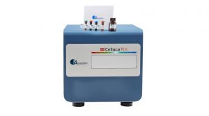

Immunophenotyping, Cell Counts, and Viability Readouts in Seconds
The Cellaca PLX system with Matrix analysis software and dedicated reagents and consumables provides a benchtop solution for accurate measurements of small sample volumes to easily perform rapid subpopulation analysis for downstream processing.
Multiplexing Made Easy
The Cellaca PLX image cytometry system is easy to use, performing simple yet sensitive cell counts, viability readouts, and multiplex analysis in seconds.
Small Sample Volume
The sample-efficient Cellaca PLX requires between 15 – 50 uL per single sample analysis. That’s 10-30 times less sample volume required per test, compared to a flow cytometer.
Speed with Sensitivity
The time to downstream processing is shortened by multiplexing with four channels with viability readouts at one minute per sample.
Optimized Assays and Kits
Our simple-to-use, mix, incubate, wash, and read reagent kits provide immunophenotyping plus viability multiplexing assays using disposable, low fluorescence consumables.
Stress-Free Software
Optimized protocols streamline surface marker staining, vitality, and apoptosis analysis with step-by-step methodologies and customizable result presentations.
Low Auto-Fluorescent Consumables
Allow for accurate surface marker detection.
Hardware, Instruments, and System Capabilities
The Cellaca PLX’s ability to consistently image, de-cluster, and report cell counts, viability, concentration, and phenotyping data makes it a versatile instrument during every stage of cell line development. The Cellaca PLX allows labs to shorten the time to downstream processing by multiplexing with up to four channels to obtain critical viability readouts at one minute per sample with no warm-up time. The cell population data can then be viewed as a histogram, scatter plot, dot plot, or contour plot for easy data viewing and analysis.
Whether using it as a complementary method for additional data or as the first stop before starting downstream assays, image cytometry can save laboratories time and prevent excessive sample loss. With this output speed, researchers can quickly perform cell purity checks at the bench and then easily continue with downstream assays without waiting for a flow cytometer. By performing multiplexing assays at the bench, laboratories can reduce the reliance on a busy flow core facility, saving flow cytometry for more complicated or detailed assays.
Expert Support for Assay Development
On-site, remote, and hybrid training are available to assist with initial setup and continued assay support for improved workflows.
Multi-Language Support
Offering over 7,000 language settings for easy global data transfer.
Remote and Field Serviceability
Calibration-free instrumentation with minimal to no routine maintenance. No clogging or liquid waste stream.
Automation Ready
Compatible with robotic integration and automated liquid handling systems.
21 CFR Part 11 Ready
- Audit trails, time stamping, user access control, and electronic signatures.
- File locking and change tracking.
- Creation of new customizable defined roles with multiple configurations available
- A close-looped database system to aid with data integrity and security.
Stress-Free Intuitive Software
- Built-in assays with optimized settings for over 400 individual cell types.
- Customizable data and calculation reports with graphs, images, charts, and tables for ease of information sharing.
The multi-color results image provides a visual of the various overlayed channels differentiating the cells while the cell count and viability data are captured below.
")
Low Auto-Fluorescent Consumables
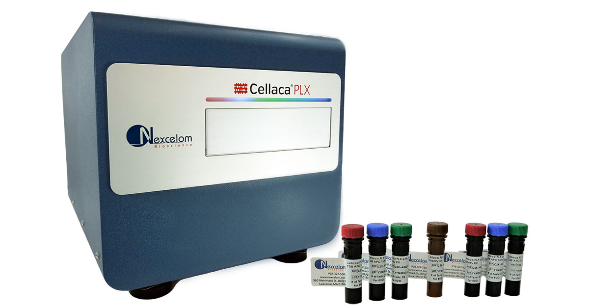
Cellaca PLX consumable slides provide a safe and self-contained environment for the sample which then does not require a decontamination procedure of the instrument at the end of each run. The slide holder, which is included with the instrument, can accommodate 4 slides or 8 samples per experimental run.
Cellaca Product Line
The Cellaca instrument line is comprised of the Cellaca MX high-throughput cell counter with brightfield and two fluorescent channels, and the Cellaca PLX image cytometry system with brightfield and five fluorescent channels for highly accurate multi-channel analysis.
The Cellaca PLX is the only multi-channel analysis instrument enabling rapid cell count, concentration, immunophenotyping, and viability readouts to significantly improve the time to downstream processing.
| Cellaca PLX | Cellaca MX FL2 | |
|---|---|---|
| Channels | Brightfield, Blue, Green, Orange, Red, Far Red | Brightfield, Green, Red |
| Number of Fluorescent Channels | 13 (6 per scan) | 3 (2 per scan) |
| Excitation LED | 370, 475, 531, 628 nm | 470, 527 nm |
| Emissions Filters | 452, 534, 605, 655, 692 nm | 534, 655 nm |
| Commonly Used Compatible Dyes and Assays | Trypan Blue, AO/PI, Hoechst, DAPI, GFP, RFP, CMFDA, Calcein AM, 7AAD, Annexin V, PE, APC, KIRAVIA Blue 520TM* | Trypan Blue, AO/PI, Calcein AM, Annexin V, Caspase 3/7 |
| Counting Speed per well | Fluorescence 4-channel scan – 1 minute | Trypan Blue – 2 seconds
Fluorescence 2-channel scan – Less than 8 seconds |
| Volume (per sample) | 15 µL in slides 50 µL – 200 µL in counting plates | 50 µL – 200 µL in counting plates |
| Size/Diameter Range | 5 – 80 µm | 5 – 80 µm |
| Concentration Range | 1×10⁵ – 1×10⁷ | 1×10⁵ – 1×10⁷ |
| IQ/OQ Option | Yes | Yes |
Associate Director of the Flow Cytometry facility at The Broad Institute, Patricia Rogers, has worked with a wide range of instrumentation and vendors in her fifteen plus years in Flow Cytometry. Patricia knows the value of great customer service and support and she believes that great instrumentation must go hand-in-hand with rapid reacting customer service to truly be exceptional. Luckily, we here at Nexcelom value the same principles and we are proud to offer excellent customer service and support to all our customers.
Watch the video to see what she says about the Cellaca MX Cell Counter.
“Anytime I have a problem you guys are always there to help me figure out a solution so you know, the service has really been more like a partnership and I’m really happy with it.”
The Cellaca PLX is Suitable for a Variety of Cell-Based Assays
Optimized Assay Reagents and Kits
The Cellaca PLX provides flow-like data with simple-to-use, mix, incubate, wash, and read reagent kits that provide immunophenotyping plus cell counts and viability multiplexing assays using disposable, low fluorescence consumables. Quickly perform cell counts, concentration, purity checks, and viability at the bench with Cellaca PLX immunophenotyping kits which combine CD3, CD4, and CD8 surface marker detection with cell viability in a single multiplex assay.
Available assay kits include:
- Immune Cell Phenotyping
- Apoptosis Detection
- High-Throughput Cell Viability
- Fluorescent Protein Analysis
| Product Name | Green | Red | Far Red | Blue | Part No. (25 tests) |
|---|---|---|---|---|---|
| 1 Surface Marker for Cell Types: PBMC, PBMC-pure, cell lines |
|||||
| KIRAVIA Blue 520™ anti-human CD3 Antibody* | CD3 | CS1-A0001-1 (25 tests) CS1-A0001-2 (100 tests) |
|||
| Cellaca® PLX, anti-human CD3 PE Antibody | CD3 | CS1-A0002-1 (25 tests) CS1-A0002-2 (100 tests) |
|||
| Cellaca® PLX, anti-human CD3 APC Antibody | CD3 | CS1-A0003-1 (25 tests) CS1-A0003-2 (100 tests) |
|||
| Cellaca® PLX, anti-human CD4 PE Antibody | CD4 | CS1-A0008-1 (25 tests) CS1-A0008-2 (100 tests) |
|||
| Cellaca® PLX, anti-human CD4 APC Antibody | CD4 | CS1-A0009-2 (100 tests) | |||
| Cellaca® PLX, anti-human CD8 PE Antibody | CD8 | CS1-A0012-1 (25 tests) CS1-A0012-2 (100 tests) |
|||
| Cellaca® PLX, anti-human CD8 APC Antibody | CD8 | CS1-A0014-1 (25 tests) CS1-A0014-2 (100 tests) |
|||
| Isotype only for Cell Types: PBMC, PBMC-pure, cell lines |
|||||
| KIRAVIA Blue 520™ Mouse IgG1, κ Isotype Ctrl Antibody* | IgG1 | CS1-A0004-1 (25 tests) CS1-A0004-2 (100 tests) |
|||
| Cellaca® PLX, PE Mouse IgG2a, κ Isotype Ctrl Antibody | IgG2a | CS1-A0005-1 (100 tests) | |||
| Cellaca® PLX, APC Mouse IgG2a, κ Isotype Ctrl Antibody | IgG2a | CS1-A0006-1 (100 tests) | |||
| Cellaca® PLX, PE Mouse IgG1, κ Isotype Ctrl (FC) Antibody | IgG1 | CS1-A0013-1 (25 tests) CS1-A0013-2 (100 tests) |
|||
| Cellaca® PLX, APC Mouse IgG1, κ Isotype Ctrl Antibody | IgG1 | CS1-A0011-1 (100 tests) | |||
| 1 Surface Markers + Viability + Isotype Controls for Cell Types: PBMC, PBMC-pure, cell lines |
|||||
| Cellaca® PLX, anti-human CD3 KB520 Viability Kit | CD3 | Dead | Total | CSK-A0001-1 (25 tests) CSK-A0001-2 (100 tests) |
|
| Cellaca® PLX, anti-human CD3 PE Viability Kit | CD3 | Dead | Total | CSK-A0002-1 (25 tests) CSK-A0002-2 (100 tests) |
|
| Cellaca® PLX, anti-human CD3 APC Viability Kit | Dead | CD3 | Total | CSK-A0003-1 (25 tests) CSK-A0003-2 (100 tests) |
|
| Cellaca® PLX, anti-human CD4 PE Viability Kit | CD4 | Dead | Total | CSK-A0004-1 (25 tests) CSK-A0004-2 (100 tests) |
|
| Cellaca® PLX, anti-human CD4 APC Viability Kit | Dead | CD4 | Total | CSK-A0005-2 (100 tests) | |
| Cellaca® PLX, anti-human CD8 PE Viability Kit | CD8 | Dead | Total | CSK-A0006-1 (25 tests) CSK-A0006-2 (100 tests) |
|
| Cellaca® PLX, anti-human CD8 APC Viability Kit | Dead | CD8 | Total | CSK-A0007-1 (25 tests) CSK-A0007-2 (100 tests) |
|
| 2 Surface Markers + Total + Isotype Controls for Cell Types: PBMCs |
|||||
| Cellaca® PLX, anti-human CD3 KB520/CD4 APC Total Cell Kit | CD3 | CD4 | Total | CSK-A0008-2 (100 tests) | |
| Cellaca® PLX, anti-human CD3 KB520/CD8 APC Total Cell Kit | CD3 | CD8 | Total | CSK-A0009-1 (25 tests) CSK-A0009-2 (100 tests) |
|
| Cellaca® PLX, anti-human CD3 APC/CD4 PE Total Cell Kit | CD4 | CD3 | Total | CSK-A0012-1 (25 tests) CSK-A0012-2 (100 tests) |
|
| Cellaca® PLX, anti-human CD3 APC/CD8 PE Total Cell Kit | CD8 | CD3 | Total | CSK-A0013-1 (25 tests) CSK-A0013-2 (100 tests) |
|
| Cellaca® PLX, anti-human CD4 PE/CD8 APC Total Cell Kit | CD4 | CD8 | Total | CSK-A0014-1 (25 tests) CSK-A0014-2 (100 tests) |
|
| 2 Surface Markers + Dead + Isotype Controls for Cell Types: PBMC-pure, cell lines |
|||||
| Cellaca® PLX, anti-human CD3 KB520/CD4 APC Dead Cell Kit | CD3 | CD4 | Dead | CSK-A0016-2 (100 tests) | |
| Cellaca® PLX, anti-human CD3 KB520/CD8 APC Dead Cell Kit | CD3 | CD8 | Dead | CSK-A0017-1 (25 tests) CSK-A0017-2 (100 tests) |
|
| Cellaca® PLX, anti-human CD3 APC/CD4 PE Dead Cell Kit | CD4 | CD3 | Dead | CSK-A0020-1 (25 tests) CSK-A0020-2 (100 tests) |
|
| Cellaca® PLX, anti-human CD3 APC/CD8 PE Dead Cell Kit | CD8 | CD3 | Dead | CSK-A0021-1 (25 tests) CSK-A0021- 2 (100 tests) |
|
| Cellaca® PLX, anti-human CD4 PE/CD8 APC Dead Cell Kit | CD4 | CD8 | Dead | CSK-A0022-1 (25 tests) CSK-A0022-2 (100 tests) |
|
| 2 Surface Markers + Viability+ Isotype Controls for Cell Types: PBMC |
|||||
| Cellaca® PLX, anti-human CD3 KB520/CD4 PE Viability Kit | CD3 | CD4 | Dead | Total | CSK-A0024-1 (25 tests) CSK-A0024-2 (100 tests) |
| Cellaca® PLX, anti-human CD3 KB520/CD8 PE Viability Kit | CD3 | CD8 | Dead | Total | CSK-A0025-1 (25 tests) CSK-A0025-2 (100 tests) |
| 3 Surface Markers + Total or Dead + Isotype Controls for Cell Types: PBMC (Total Cell Kit Only) for Cell Types: PBMC-pure, cell lines (Dead Cell Kit Only) |
|||||
| Cellaca® PLX, anti-human CD3 KB520/CD4 PE/CD8 APC Total Cell Kit | CD3 | CD4 | CD8 | Total | CSK-A0026-1 (25 tests) CSK-A0026-2 (100 tests) |
| Cellaca® PLX, anti-human CD3 KB520/CD4 PE/CD8 APC Dead Cell Kit | CD3 | CD4 | CD8 | Dead | CSK-A0027-1 (25 tests) CSK-A0027-2 (100 tests) |
| For Apoptosis for Cell Types: cell lines |
|||||
| Cellaca® PLX, Caspase 3/RubyDead Apoptosis Kit | Casp | Dead | Total | CSK-A0028-2 (100 tests) | |
| Cellaca® PLX, Annexin V-FITC/PI Apoptosis Kit | AV | PI | Total | CSK-A0029-1 (25 tests) CSK-A0029-2 (100 tests) |
|
| For Fluorescent Proteins for Cell Types: cell lines |
|||||
| Cellaca® PLX, Hoechst/RubyDead Viability Kit | Dead | Total | CSK-A0030-1 (25 tests) CSK-A0030-2 (100 tests) |
||
| Slides for Cell Types: PBMC, PBMC-pure, cell lines |
|||||
| Cellaca® PLX, Low Fluorescence Slides | CHM2-ACR-001 (100 tests) CHM2-ACR-002 (500 tests) |
||||
*KIRAVIA Blue 520™ is a trademark of Sony. This product is subject to proprietary rights of Sony and is made and sold under license from Sony Corporation.
Associate Director of the Flow Cytometry facility at The Broad Institute, Patricia Rogers, has worked with a wide range of instrumentation and vendors in her fifteen plus years in Flow Cytometry. Patricia knows the value of great customer service and support and she believes that great instrumentation must go hand-in-hand with rapid reacting customer service to truly be exceptional. Luckily, we here at Nexcelom value the same principles and we are proud to offer excellent customer service and support to all our customers.
Watch the video to see what she says about the Cellaca MX Cell Counter.
“Anytime I have a problem you guys are always there to help me figure out a solution so you know, the service has really been more like a partnership and I’m really happy with it.”
The Cellaca PLX Applications
Download the Application Note: Novel High-Throughput Image Cytometry Methods for T-Cell Immunophenotyping and Viability Readouts
Eliminate Complex Procedures and Precious Sample Loss
With the Cellaca PLX Platform, you can:
- Quickly perform cell purity checks and viability at the bench with Cellaca PLX immunophenotyping kits which combine CD3, CD4, and CD8 surface marker detection with cell viability in a single multiplex assay.
- Stain transfected cell lines with viability dyes to determine the percentage of transfected or transduced cells that are live/dead.
- Perform routine viability measurements as well as apoptosis functional assays to determine cell health.
Immunophenotyping Application:
Routine CD3, CD4, and CD8 population analysis is a standard practice in cell and gene therapy research. Understanding specific cell populations and viability allows researchers to move tested samples to subsequent downstream assays.
A. Brightfield

B. Hoechst

C. CD3-KIRAVIA Blue 520™

D. CD4-PE

E. CD8-APC

Surface marker stained PBMC sample
PBMCs were stained with CD3, CD4, and CD8 surface markers, along with the total dye Hoechst. Image data (A-E) from the Cellaca PLX was exported to FCS Express™ for analysis. Gated nucleated (Hoechst +) CD3+ cells (~70% of total cells) are shown in the first scatter plot (F). The CD3 positive population was then further gated for CD4+ (~62%) and CD8+ (~28%) positive cells (G).
F. Hoechst+, CD3+

G. CD4+, CD8+

Apoptosis Application:
Apoptosis, viability, and other cell health indicators are vital in examining the state of collected patient samples. When sample viability drops below the set benchmark researchers often conduct multiple assays, including apoptosis, to understand possible causes for cell viability decline.
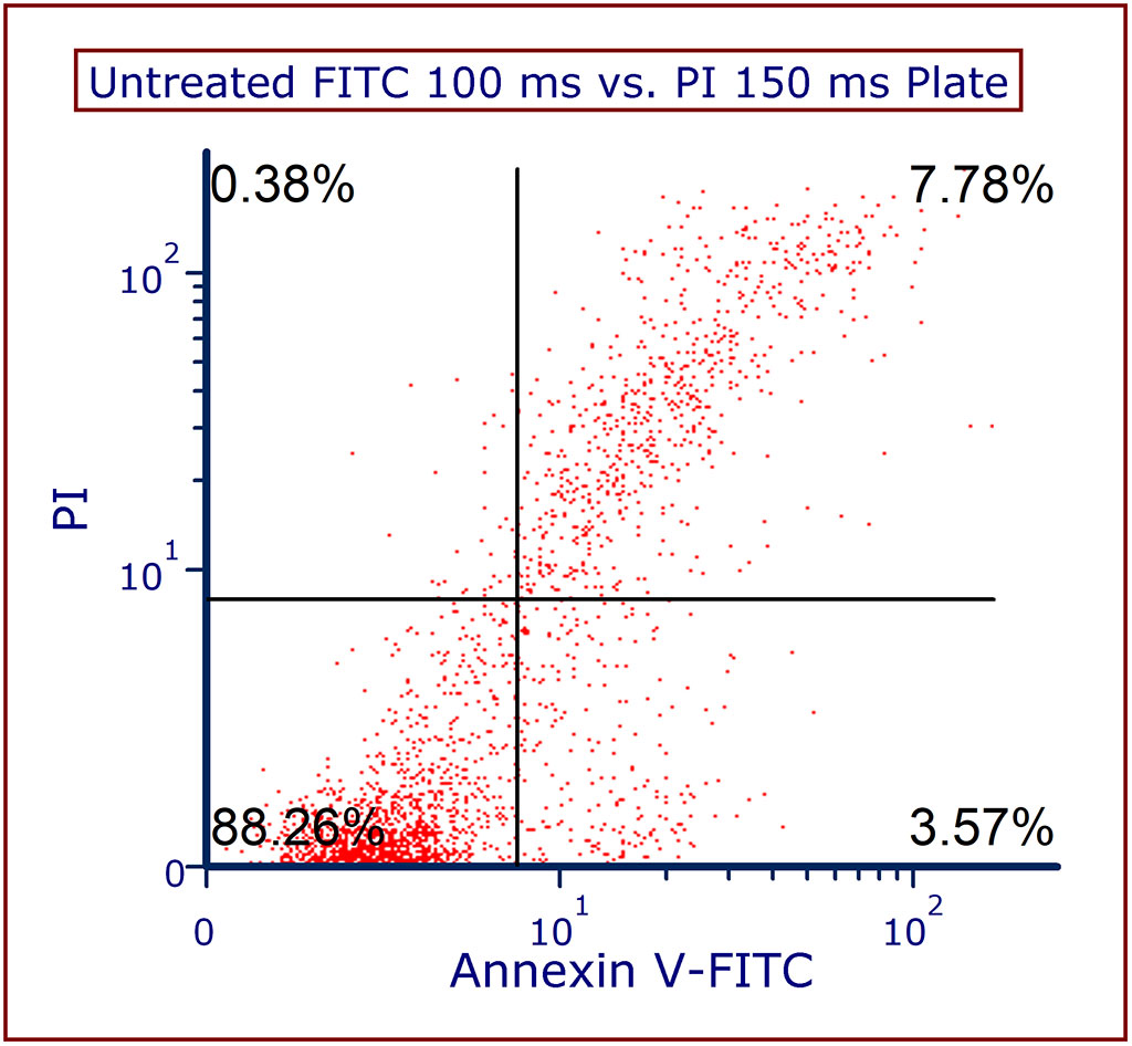
Exported image data in this scatter plot is displaying Jurkat cells stained with annexin V-FITC and Propidium iodide. Healthy annexin V and PI negative cells (88%) are in the left bottom quadrant.
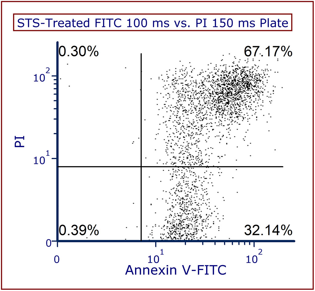
Jurkat cells were treated with staurosporine (STS), imaged on the Cellaca PLX, and data exported into FCS Express™. Compared to the healthy control, there was a significant increase in annexin V positive cells from 3.5% to 32%. The number of double positive cells for annexin V and PI also drastically increased from 7.7% to 67%.
Fluorescent Protein Expression Application:
Transduction and transfection assays are often performed not only in cell and gene therapy but also in cell line development, viral vaccines, and multiple other areas of research. Quantifying the expression of fluorescent proteins, such as GFP and RFP, and the viability of those cells, is essential in determining whether gene insertion was a success.
")
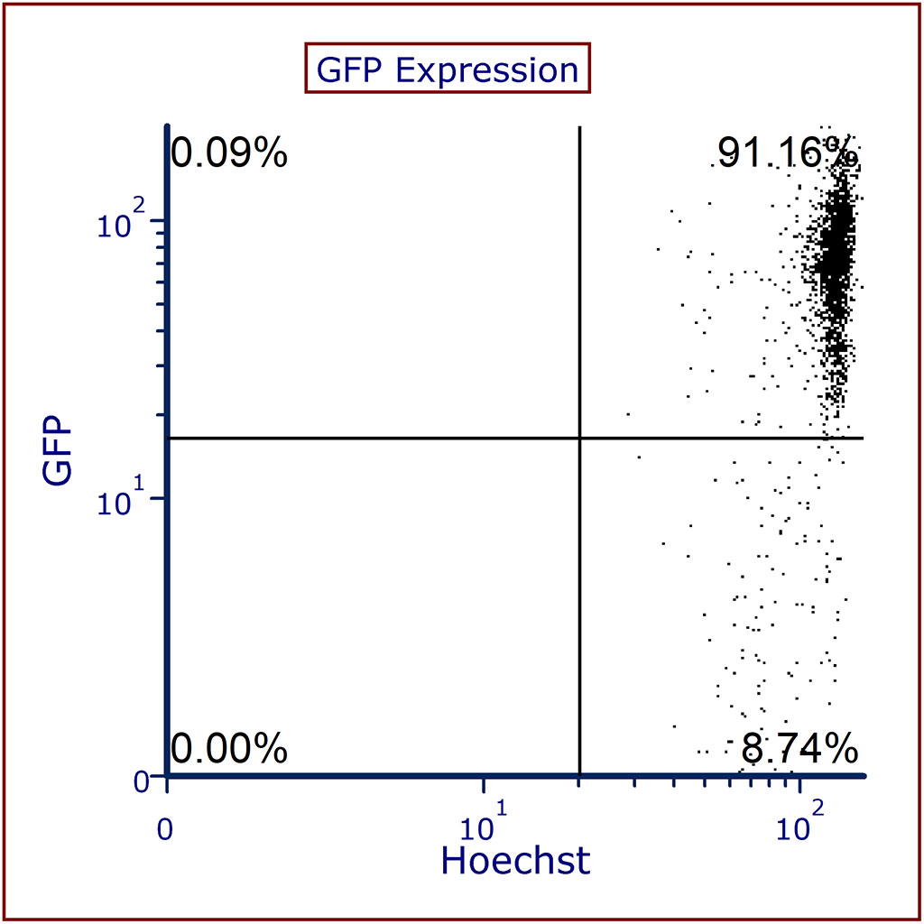
The scatter plot shows GFP expressing cells counterstained with Hoechst. The top right quadrant is showing that 91% of cells are nucleated GFP positive cells while 8% of the population are nucleated cells that are GFP negative.
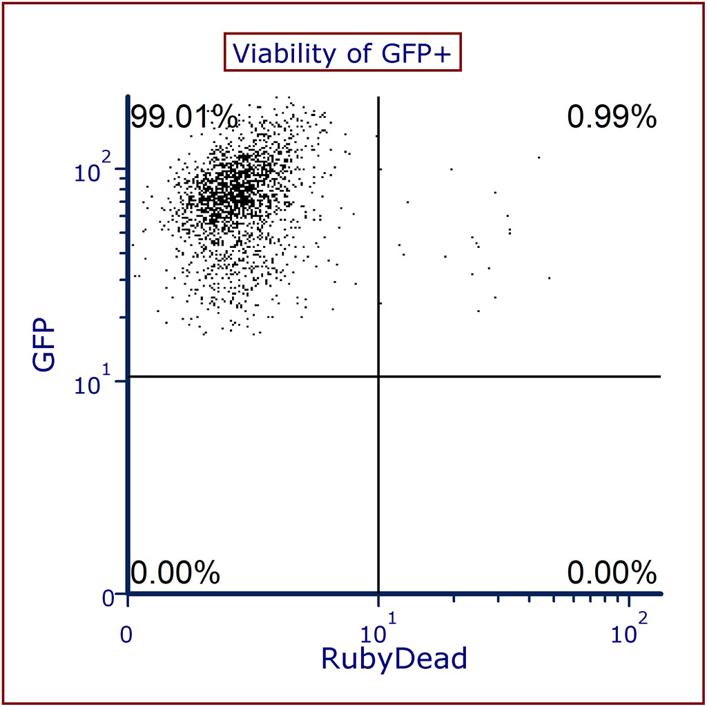
GFP in the scatter plot shows GFP-expressing cells stained with a viability dye, RubyDead. The population of cells in the top left quadrant shows that nearly all (99%) of GFP-expressing cells one live.
Associate Director of the Flow Cytometry facility at The Broad Institute, Patricia Rogers, has worked with a wide range of instrumentation and vendors in her fifteen plus years in Flow Cytometry. Patricia knows the value of great customer service and support and she believes that great instrumentation must go hand-in-hand with rapid reacting customer service to truly be exceptional. Luckily, we here at Nexcelom value the same principles and we are proud to offer excellent customer service and support to all our customers.
Watch the video to see what she says about the Cellaca MX Cell Counter.
“Anytime I have a problem you guys are always there to help me figure out a solution so you know, the service has really been more like a partnership and I’m really happy with it.”