
Celigo Image Cytometer
Whole-well image cytometer for adherent and suspension cells
See how the Celigo Imaging Cytometer works »
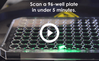
Celigo plate scan speed
See the Celigo key features
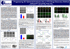
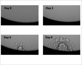
Featured Poster: Progressing 3D Spheroid Analysis into a HTS Drug Discovery Method

Introduction to Celigo
Celigo is a plate-based benchtop brightfield and fluorescent imaging system designed for whole-well live-cell analysis and cell sample characterization. Celigo images and analyzes cells in various types of vessels including 6 – 1536 well plates, T25, T75 flasks, 10 cm dishes, and glass slides without disturbing their natural state.
Individual cell level analysis is easily generated, providing cell level insights unlike ELISA or protein-based assays, and at a faster rate than flow cytometry. A broad range of complex cell-based assays have been optimized for Celigo:
Apoptosis | Cell Cycle | Fluorescent Reporters | Cytotoxicity | Label-free Proliferation
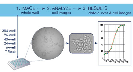
Rapid Whole-well Imaging and Analysis
- Sensitive whole-well imaging ensures accurate cell population analysis
- Remove non-uniform seeding from the equation
- Accurately detect, image, and count every cell in every well
- Unique scanning mirrors enable fast, high-quality images

Best-in-Class Brightfield and Four Fluorescent Channels
- Proprietary algorithm for simultaneous imaging and label-free cell analysis
- Perform real-time multiplex assays with four fluorescent channels
Convenient Workflow Designed for Biologists
- Multiple object-driven algorithms and a variety of assay-based applications
- Accessible adaptation with easy-to-follow software
- Save customizable experiments as well as default settings
- Built-in gating parameters allow easy data analysis and visualization
- Real-time graphic feedback allows multiple parameters to be measured simultaneously for intuitive analysis:
- Cell growth | Viability | Morphology | Count | Concentration
Automation and Data Management
- Accessible application programming interface (API) for easy automated workflow integration
- Automated microplate handling for kinetic end-point analysis or time-point analysis
“The Celigo has increased our research output and has dramatically sped up our discovery process because of its ease and simplicity of use. It is a wonderful tool for our institution to have.”
“The aspects of being able to image proliferation are key to our experiments. The Celigo Imaging Cytometer is very user friendly and the support staff is outstanding!”
Count Every Cell in Every Well
Perform best-in-class brightfield imaging in combination with four fluorescence channels to provide high speed, fully automated imaging and quantification of suspension AND adherent cells, tumor spheroids, iPSC, and cancer stem cell colonies.
Proprietary optics and scanning system enable fast imaging of the entire well while maintaining consistent illumination and contrast out to the well edge, for accurate identification of all cells within each well.
With an intuitive software interface and optional integration into automation platforms, the Celigo provides labs with increased capabilities. Celigo is valuable at every step from enabling high-throughput screens of drug libraries in early drug discovery or running high-throughput validated assays for late-stage research.
Best-in-Class Brightfield Imaging for All Well Sizes
Excellent Optics for Enhanced Image Quality
Improves brightfield optical image quality at the edge of wells and eliminates edge optical distortion by using an F-Theta lens for superior well edge-to-edge image contrast.
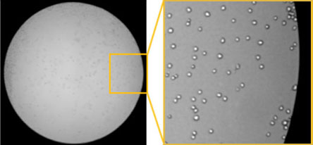
Celigo Imaging Cytometer brightfield image showing the edge of a well on a 96-well microplate showing enhanced image quality and contrast.
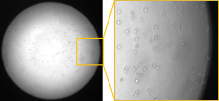
Conventional microscope brightfield image showing distortion and low contrast near the edge of the field-of-view.
Fast Plate Scanning for Image Acquisition and Analysis
Increase plate-acquisition speed using novel plate scanning technology for fast acquisition. Scan a 384-well plate in brightfield in two minutes or less.
Ability to Scan a Variety of Plate Vessels
Scan wells of any size using automated image stitching that can view and quantify cells and colonies in vessels up to 6-well plates and 10 cm dishes.
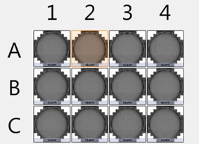
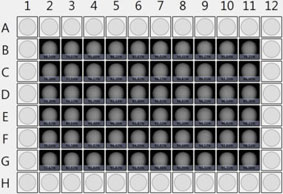
Examples of a 12-well and 96-well plate acquisition using Celigo Imaging Cytometer.
Accurately Quantify Cells and Colonies
Accurately quantify every cell and colony in the well even if they do not grow uniformly across a well.
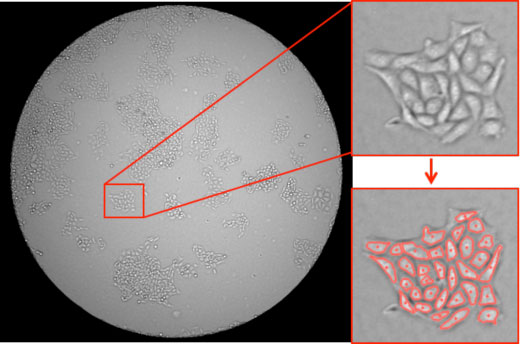
Brightfield image of a well on a 96-well microplate showing counted cell colonies across the well.
Accurately Measure Adherent Cells without Trypsinization
Analyze your cell sample without trypsinization to avoid losing cells and look at cells right where they grow over multiple scan times.
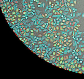
Brightfield image of a well on a 96-well microplate showing counted adherent cells.
Brightfield and Four Fluorescent Channels (UV to Far Red) for Analyzing & Quantifying Multiplex Assays
Brightfield and Multi-Channel Fluorescence Imaging
Use a combination of brightfield and fluorescent imaging allowing the development of multicolor assays ranging from UV to Far Red.
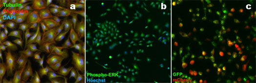
Fluorescent images acquired with Celigo Imaging Cytometer of (a) Tubulin, Phalloidin, and DAPI, (b) Phospho-ERK and Hoechst, and (c) GFP and mCherry.
Clear Visualization of Image and Data Correlation
Correlate the cell populations identified in gates with the cells visualized in the image using color-coding overlays.
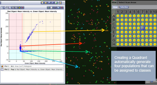
Correlation of gated scatter plot data to counted cells on acquired images.
Powerful Image Analysis Software
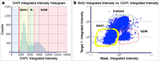
Flow-like data analysis and presentation generated by Celigo Imaging Cytometer. Fluorescence intensity (a) histogram and (b) scatter plot.
Convenient Data Visualization Environment that Facilitates Data Interpretation
Simple At-a-Glance Plate-Based Data Review and Growth Curves
Quickly identify cells from well to well, easily evaluate data, and generate time course growth curves to be easily displayed in a plate overview for at-a-glance data review for each plate.
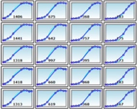
384-well plate view of wells showing cell confluency on the plate. Time-course data plot generated by Celigo Imaging Cytometer.
Easy Brightfield and Fluorescent Image Navigation
Navigate between plates, wells, and time points to look at your cells growing in specific well locations.
Brightfield image of a single cell colony observed at the same location of a well on a 96-well plate.
Application Programming Interface (API) and Data Management Solution for Integration into Automated Workflows
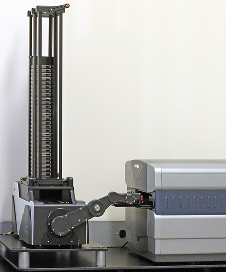
Integrate with Robotics
Automate your assays by running the Celigo under the control of scheduling software and integrate with robotic arms, plate stackers, automated incubators, and liquid handlers.
Automated System for High-throughput Integrated Data Acquisition and Analysis
Acquire images and data analysis of hundreds of plates 24/7 automatically.
Time-saving Data Processing Method
Get data on the fly by acquiring and analyzing images simultaneously.
Flexible Data Analysis Method for Multiple Users
Analyze large data sets off the Celigo instrument using the Celigo Satellite Workstation and free up Celigo time to acquire more plates.
Series of Customized Applications for Each Assay – Ready to Use, Does Not Require Any Image Analysis Expertise
Label-Free Brightfield Cell Analysis
Take advantage of label-free brightfield applications to avoid staining cells with toxic dyes or transfecting with fluorescent reporters.
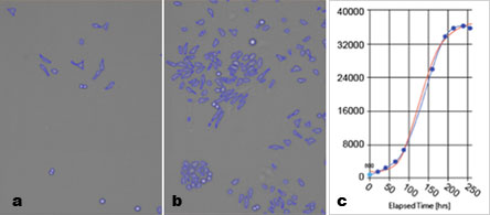
Label-free brightfield cell counting and from (a) low count to (b) high cell count which can be used to directly generate (c) a cell growth curve
Numerous Cell Characterization Assays
Live cell analysis of images for cell counting, confluence, colonies, and 3D-spheroids.
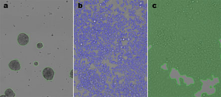
Label-free brightfield cell counting of (a) spheroids, (b) cell counting, or (c) confluence percentage.
Ability to Export Brightfield and Fluorescent Images for Publications
Capitalize on the Celigo brightfield and fluorescent image quality to take images of your cells for your records and strengthen your publications.
Easily Access and Manage Data using Celigo Network Database
Facilitate access to your imaging data using the Celigo network database and connect multiple instruments and satellite workstations to seamlessly acquire and analyze your data from a convenient centralized location.
Easy-to-Use Preset Assay Parameters for Quick Data Analysis
Perform Celigo assays using pre-defined parameters that require very few modifications from the researchers.
Simple and Intuitive Software User Interface
Benefit from a simple and intuitive four-step software workflow to run all your assays.
Celigo Express with 21 CFR Part 11 Module
Celigo Express offers a 21 CFR Part 11 module that is compliant with the Code of Federal Regulations (CFR) Title 21 Part 11 − Electronic Records: Electronic Signatures published by the U. S. Food and Drug Administration (FDA). This module ensures that an organization’s use of electronic records and digital signatures in place of traditional paper-based documentation complies with current FDA regulations.
The newly developed Celigo Express software works with the 21 CFR Part 11 ready module to simplify the data acquisition process by allowing researchers to use projects imported from Celigo Pro (v5.4) and quickly run them to collect sample data. With a few clicks of the mouse, run a scan using Celigo Express:
- Select the project to run
- Click on Start Scan
- View your data

Key elements of the 21 CFR Part 11 module are:
- Electronic Signatures are captured during the counting and analysis workflow using e-Sign/e-Unsign buttons and include the name of an authorized user, the date/time when the signature was executed, and the meaning of the signature (i.e., indicating either Approval or Rejection of an action).
- An Audit Trail assures the integrity of an electronic record and continually monitors all users performing actions, the type of actions being performed, and the date/time associated with user actions.
- User Roles allows for specialized access control to the varying levels of Celigo Express functionality to specified groups. When creating a role, subsets of privileges can be selected to apply to users performing common or specialized tasks.
- A Close-looped Database System ensures the integrity of the data generated.
Understanding User Role Privileges
- Data − Controls whether users can create, delete, export/import, and approve/reject data during the counting and analysis workflow.
- Settings − Controls whether users can access the Projects screen functionality available under the Manage tab to allow them to update, delete, and import/export projects.
- Users, Roles, or Audits − Controls whether users can access the 21 CFR Part 11 module Users, Roles, or Audits options under the Manage tab to allow them to create, update, and delete Celigo Express users/roles, or monitor and export the audit trail automatically generated by the system after the module is enabled.

Expand Your Lab’s Image Analysis & Data Management Capabilities
Storing, managing and sharing large volumes of image data can be a challenge. That’s why we’ve made the Signals Image Artist™ image analysis and management platform* from Revvity compatible with Celigo image data for use alongside the Celigo system’s own powerful acquisition, visualization and analysis software.
With the addition of the Signals Image Artist platform to your lab, you’ll be able to quickly process, store, and share all your image data from the Celigo image cytometer and other major cell imaging systems in a single central location. Its powerful image processing capabilities and ready-made analysis building blocks will provide you with more options for exploring your data and gaining new insights. Benefit from machine learning and artificial intelligence (AI) capabilities to train the software to develop analysis algorithms – and get answers faster. You can even access data and perform analysis remotely via the browser login.
- Compatible with Celigo image cytometer and major cell imaging systems
- A central location to store and access all your image data – from the lab, office, or remotely
- Advanced image analysis capabilities, including machine learning and AI
- Multi-user solution that can support your entire lab without compromising performance
- Scalable data storage to expand with your labs needs over time
- Easy IT management with single system and installation across image data types
- Cloud and on-premise options to choose from
- Web browser application to access data and perform analysis remotely
Contact us via the request form for more details.
*Signals Image Artist platform is sold separately – it is not sold as part of the Celigo system.

Monitoring the effects of a drug panel on invasion of U87MG Glioblastoma MCTS into Basement Membrane Extract (BME) Matrigel. Signals Image Artist’s “Find Image Region” building block is used on the brightfield channel to segment the membrane region of spheroids.
Take Your Analysis to the Next Level, Automation!
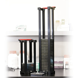
Stacker Automation: No Need to ‘Baby-Sit’ Your Plates
With the simple integration of the plate stacker, you are free to work on other tasks while Celigo screens your stack of plates.
- Up to 50 plate capacity
- 15-sec transfer rate between plates
- Data exports automatically for each plate
- Handles plates with or without lids
- Accommodates from 6-well up to 1536-well plate formats
- Fits on a standard lab bench
- Ideal for endpoint assays
- Easy to add on to existing Celigo instruments
Video: Watch the Celigo with integrated Plate Stacker
| Parameter | Specification |
|---|---|
| Celigo only Dimensions | 19 inch wide x 29 inch long x 17 inch high (with plate holder extended) |
| Celigo + Stacker Dimensions | 25 inch wide x 44 inch long x 40 inch high (Total) |
| Electrical Power | 5 Electrical outlets needed at 100-240VAC 50/60Hz (30Amps Total) |
| Compressor (sold with Stacker) | 1.2 CFM @ 90 PSI with 1 gallon tank |
| Operating Temperature | 15 ºC to 25 ºC |
| Operating Humidity | 10% to 90% RH, non-condensing |
| Instrument Shipping & Storage Temperature | -18 ºC to 65 ºC |
| Instrument Shipping & Storage Humidity | 10% to 90% RH, non-condensing |
| Manufacturer | Nexcelom Bioscience, LLC. 360 Merrimack St., Building 9 Lawrence, MA 01843Kbiosystems Limited Units 5 to 10 Paycocke Close Basildon, Essex, UK, SS143HS |
| Distributor/Support | Obtain information at https://www.nexcelom.com/purchase or contact support@nexcelom.com |
Applications
Imaging Vessels
| Plate Name | Manufacturer | Well Type | Compatibles |
|---|---|---|---|
| 6-Well BD Falcon™ 353046 Plate | Corning | Clear | 353224, 353934, 353846, 351146, 353502 |
| 6-Well Corning™ 3516 Plate | Corning | Clear | 3471, 3506, 3335 |
| 6-Well CytoOne® CC7682-7506 Plate | CytoOne | Clear | — |
| 6-Well Greiner™ 657160 Plate | Greiner | Clear | 657185, 657165 |
| 6-Well Nunc™ 140675 Plate | Thermo | Clear | — |
| 12-Well BD Falcon™ 353043 Plate | Corning | Clear | 353224, 351143, 353503 |
| 12-Well Corning™ 3513 Plate | Corning | Clear | 3336, 3512 |
| 12-Well CytoOne® CC7682-7512 Plate | CytoOne | Clear | — |
| 24-Well BD Falcon 353047 Plate | Corning | Clear | 353226, 353935, 353847, 351147, 358115,354723, 356723, 354775, 356775, 353504 |
| 24-Well Corning 3524 Plate | Corning | Clear | 3337, 3526, 3527, 3573 |
| 24-Well CytoOne CC7682-7524 Plate | CytoOne | Clear | — |
| 24-Well Greiner 662160 Plate | Greiner | Clear | 662102, 622165 |
| 24-Well Visiplate 1450606 Plate | Revvity | Black | — |
| 24-Well Seahorse XF24 Plate | Seahorse Biosciences | Clear | — |
| 48-Well Corning 3548 Plate | Corning | Clear | — |
| 48-Well Greiner 677180 Plate | Greiner | Clear | 677102, 677165 |
| 96-Well BD Falcon 353219 Plate | Corning | Black, White | 353377 |
| 96-Well BD Falcon 354640 Plate | Corning | Black, White | 354650, 356650, 354651, 356651, 356701, 356693, 354649, 356649, 356640, 356700, 356692, 356717 |
| 96-Well BD Falcon 356717 Plate | Corning | Black | 354717 |
| 96-Well BD Falcon 351177 U-Bottom Plate | Corning | Clear, Round Bottom ULA | — |
| 96-Well BD Falcon 353072 Plate | Corning | Clear | 353916, 353936, 353075, 351172, 354407, 354429, 354461, 356461, 354516, 356516, 354607, 356698, 356690, 354689, 356689, 354409, 354410, 354670, 354596, 354657 |
| 96-Well BD Falcon 353219 Plate | Corning | Black, White | 353377 |
| 96-Well BD Falcon 354640 Plate | Corning | Black, White | 354650, 356650, 354651, 356651, 356701, 356693, 354649, 356649, 356640, 356700, 356692, 356717 |
| 96-Well BD Falcon 356717 Plate | Corning | Black | 354717 |
| 96-Well BD Falcon 351177 U-Bottom Plate | Corning | Clear, Round Bottom | — |
| 96-Well Corning 3596 Plate | Corning | Clear | 3300, 3474, 3595, 3598, 3599, 3585, 3595, 3628, 3841 |
| 96-Well Corning 3596 Plate | Corning | Clear | 3300, 3474, 3595, 3598, 3599, 3585, 3595, 3628, 3841 |
| 96-Well Corning 3603 Plate | Corning | Black, White | 3604, 3610, 3631, 3632, 3651, 3843, 3842, 3903, 3904, 3601, 3635, 3340, 3842, 3843 |
| 96-Well Corning 3696 Plate | Corning | Half Area, Black | 3686, 3688, 3690, 3693, 3694, 3695, 3696, 3697 |
| 96-Well Corning 7007 U-Bottom Plate | Corning | Clear, Round Bottom | 3366, 3797, 3360, 3367, 3788, 3795, 3798, 3605, 3789, 3792, 3799 |
| 96-Well Greiner 655090 Plate | Greiner | Black, White | 655087, 655097, 655946, 655948, 655936, 655956, 655098, 655094, 655944 |
| 96-Well Greiner 655087 Plate | Greiner | Black | 655088 |
| 96-Well Greiner 655161 Plate | Greiner | Clear | 655101, 655192 |
| 96-Well Greiner 655180 Plate | Greiner | Clear, chimney | 655182, 655185, 655940, 655930, 655950 |
| 96-Well Greiner 675986 Plate | Greiner | Half Area, Black | 67509x |
| 96-Well Greiner 650185 U-Bottom Plate | Greiner | Clear, Round Bottom ULA | — |
| 96-well ULA-96U Plate | Revvity | Clear, Round Bottom, ULA | — |
| 96-Well Nunc 167008 Plate | Thermo | Clear | — |
| 96-Well Viewplate 6005225 Plate | Revvity | Black | — |
| 96-Well Isoplate 6005050 Plate | Revvity | Black | — |
| 96-Well Seahorse FX96 Plate | Seahorse Bioscience | Black | — |
| 384-well ULA-384U Plate | Revvity | Clear, Round Bottom, ULA | — |
| 384-Well BD Falcon 353962 Plate | Corning | Clear | — |
| 384-Well Corning 3542 Plate | Corning | Low volume, Black | 3540 |
| 384-Well Corning 3680 Plate | Corning | Clear | 3640, 3844, 3700, 3701, 3702, 3844 |
| 384-Well Corning 3712 Plate | Corning | Black, White | 3653, 3655, 3846, 3845, 3706, 3707, 3711, 3683, 3845, 3846 |
| 384-Well Corning 3827 Plate | Corning | Low attach | — |
| 384-Well Greiner 781182 Plate | Greiner | Clear | 781185, 781186, 781061, 781940, 781930, 781950 |
| 384-Well Greiner 781091 Plate | Greiner | Black | 781098, 781095, 781094, 781944, 781090, 781096, 781097, 781946, 781948, 781936, 781956 |
| 1536-Well Corning 3838 Plate | Corning | Black, White | 3833, 3836, 3893 |
| 1536-Well BD Falcon 356771 Plate | Corning | Black | — |
| 1536-Well Greiner 789866 Plate | Greiner | Black | 789896 |
| 1-Well Nunc Omnitray | Thermo Fisher | Clear | 242811 |
| T25 Greiner 690175 Flask | Greiner | Clear | — |
| T25 Greiner 690175 Flask – Single View | Greiner | Clear | — |
| T25 BD Falcon 353014 Flask | Corning | Clear | — |
| T25 BD Falcon 353014 Flask – Single View | Corning | Clear | — |
| T75 BD Falcon 353136 Flask | Corning | Clear | — |
| T75 BD Falcon 353136 Flask – Single View | Corning | Clear | — |
| 10cm Dish BD Falcon 353003 Dish | Corning | Clear | 353803 |
| 1-Slide Holder (2/3 cover slip) | Revvity | Clear | — |
| 1-Slide Holder (square cover slip) | Revvity | Clear | — |
| 4-Slide Holder (2/3 cover slip) | Revvity | Clear | — |
| 4-Slide Holder (square cover slip) | Revvity | Clear | — |
Celigo Specifications
Software
- Proprietary image acquisition and processing software
- Powerful analysis software/Dell Precision computer
- Windows 10
Illumination/Optics
- 1 LED-based enhanced brightfield imaging channel with uniform well illumination
- 4 LED-based fluorescent channels
- Proprietary F-theta lens with superior well edge-to-edge contrast
- Galvanometric mirrors for fast imaging of large areas
- Large chip CCD camera (2024 x 2024 pixels)
- 1, 2, 4 or 8 μm/pixel resolution
Fluorescent Channels
| Channel | Excitation | Dichroic | Emission | Typical Dyes |
|---|---|---|---|---|
| Blue | 377/50 | 409 | 470/22 | Hoechst, DAPI |
| Green | 483/32 | 506 | 536/40 | FITC, Calcein, GFP, AlexaFluor® 488 |
| Red | 531/40 | 593 | 629/53 | R-PE, PI, Texas Red, AlexaFluor® 568 |
| Far-Red | 628/40 | 660 | 688/31 | DRAQ5®, AlexaFluor® 647 |
Plate Compatibility
- 6, 12, 24 48, 96, 384, 1536 well plates (black, white and clear wall plates)
- T-25 and T-75 flasks
- Slides and cell arrays plate profiles available upon request
High-Speed Imaging
- Less than 2 minutes per 384-well plate
Weight and Dimensions
- Dimensions: 19.5″W x 16″H x 24″D (49.5cm x 40cm x 61cm)
- Weight: 117 lbs. (53 kg)
Power Requirements
- 110-220 VAC 50-60 Hz
Regulatory Compliance
- CE marking
Celigo Products
Celigo S Imaging Cytometer
Celigo S Brightfield Only
Celigo S with Automation License
Celigo S Brightfield Only with Automation License
Celigo S Satellite Workstation
Celigo Imaging Cytometer – 5 Channels
Celigo Imaging Cytometer – 5 Channels with Automation License
Part Numbers
200-BFFL-S
200-BF-S
200-BFFL-S-AUTO
200-BF-S-AUTO
200-CSW-S
200-BFFL-5C
200-BFFL-5C-AUTO
Celigo Service & Upgrades
Celigo Automation Upgrade
Celigo S Upgrade
Celigo Fluorescent Upgrade
Celigo Software Upgrade
Celigo Service Contract
Celigo Brightfield Only Service Contract
Part Numbers
200-AUTO-UPG
200-S-UPG
200-FL-UPG
Accessories
Celigo 1 Slide Holder
Celigo 4 Slides Holder
Celigo 10 cm Dish Holder
Part Numbers
200-1SL-HLDR
200-4SL-HLDR
200-10CM-HLDR
Reagents
| Application | Product Name | Description | Product Number |
|---|---|---|---|
| Apoptosis | ViaStain™ Live Caspase 3/7 Detection for 2D/3D Culture | Measure apoptosis in 2D and 3D cultures to perform both real-time kinetic and end point assays. | CS1-V0002-1 |
| Apoptosis | ViaStain™ Live Caspase 3/7 Detection for 2D/3D Culture with Hoechst | Measure apoptosis in 2D and 3D cultures for end point assays with Hoechst staining. | CSK-V0003-1 |
| Apoptosis | ViaStain™ No-Wash Annexin V-FITC Kit for Celigo | A no-wash assay designed to discriminate between healthy, apoptotic, dead cells by simultaneously staining the cells with Annexin V, propidium iodide (PI) and Hoechst | CSK-V0007-1 |
| Proliferation/Tracer | ViaStain™ CFSE | A green cell tracer dye used for labeling live cells and monitoring proliferation over multiple generations. | CS1-P0002-1 |
| Proliferation/Tracer | ViaStain™ CMFDA | A green cell tracer dye used for labeling live cells and is retained in the cells over several generations. | CS1-P0001-1 |
| Tracer | ViaStain™ Tracer Blue | A dye used for labeling live cells. Once it enters the cells, the blue color dye is retained inside live cells. | CS1-P0003-1 |
| Tracer | ViaStain™ Dead Cell Nuclear Blue | Bright blue dye designed to detect dead nucleated cells in an end point assay. No wash step, add and read assay. | CSK-V0015-1 |
| Tracer | ViaStain™ Dead Cell Nuclear Far Red | Bright far red dye designed to detect dead nucleated cells in an end point assay. No wash step, add and read assay. | CSK-V0014-1 |
| Tracer | ViaStain™ Dead Cell Nuclear Green | Bright green dye designed to detect dead nucleated cells in an end point assay. No wash step, add and read assay. | CSK-V0012-1 |
| Tracer | ViaStain™ Dead Cell Nuclear Red | Bright red dye designed to detect dead nucleated cells in an end point assay. No wash step, add and read assay. | CSK-V0013-1 |
| Tracer | ViaStain™ Total Cell Nuclear Far Red | Bright blue dye designed to detect nucleated cells in an end point assay. No wash step, add and read assay. | CSK-V0010-1 |
| Tracer | ViaStain™ Total Cell Nuclear Green | Bright green dye designed to detect nucleated cells in an end point assay. No wash step, add and read assay. | CSK-V0008-1 |
| Tracer | ViaStain™ Total Cell Nuclear Red | Bright red dye designed to detect nucleated cells in an end point assay. No wash step, add and read assay. | CSK-V0009-1 |
| Viability | DAPI | A fluorescent dye that binds to DNA. Excited by UV light, and may be used to stain fixed cells. | CS1-0127-2mL |
| Viability | Hoechst 33342 | A fluorescent dye, excited by UV light, that binds to DNA. More cell membrane permeable than other Hoechst dyes, and can be used to stain live cells. | CS1-0128-5mL |
| Viability | ViaStain™ Calcein AM/Hoechst/PI Viability kit | Live and metabolically active cells are labled with Calcein, dead cells are labeled with PI, and total number of cells is determined by Hoechst. | CSK-V0006-1 |
| Viability | ViaStain™ Hoechst/PI | A no wash assay kit designed to determine cell viability by staining dead cells with PI and all cell with Hoechst 33342 | CSK-V0005-1 |
| Viability | ViaStain™ PI Staining Solution | Propidium Iodide: a membrane impermeable staining solution for the staining of dead nucleated cells. | CS1-0109-5mL |
| Viability | ViaStain™ AO Staining Solution | Acridine Orange: a membrane permeable staining solution for the detection of nucleated cells. | CS1-0108-5mL |
| Viability | ViaStain™ AOPI Staining Solution | A staining solution for the detection of live and dead nucleated mammalian cells. | CS2-0106-5mL CS2-0106-25mL |
Resources
Manuals
Literature
- Celigo Brochure
- Celigo Applications Guide
- Celigo Plate Profiles
- Signals Image Artist for Celigo Flyer
Videos
- Demo on Demand: Modern Virology Cell-based Assays on the Celigo Image Cytometer
- Demo on Demand: Modern Cell-based Assays for Cell and Gene Therapy Using Image Cytometry
Scientific Posters
- A rapid high-throughput 3D tumor spheroid image cytometry screening method for drug discovery
- Imaging and Analysis of 3D Patient-Derived Organoids Using the Celigo Automated Image Cytometer
- Real-time kinetic viability and apoptosis detection of 3D multicellular tumor spheroids using the Image Cytometer
- Automated high-throughput method for assessing pathogenic infectious dose (TCID50) using Celigo imaging cytometer
- Novel cell-based high-throughput hybridoma screening method using the Celigo image cytometer for antibody discovery
- Validating and optimizing single cell sorting of FACS using Celigo image cytometry
- High-throughput detection of CRISPR/Cas 9 gene editing efficiency, cell proliferation/viability, and monoclonality validation using Celigo Image Cytometer
- Automation Method to Increase Efficiency in Cell Line Development
- High-throughput method to analyze the cytotoxicity of CAR-T Cells in a 3D tumor spheroid model using image cytometry
Application Notes
- Automated label free growth tracking and culture management within flasks and multi well plates
- High-throughput counting of crystal violet stained plaques
- Viral titration assay using adherent cells
- Kinetic apoptosis using Caspase 3/7
- Celigo provides an alternative method to Cell Titer-Glo for proliferation studies in suspension cells
- Normalization of Agilent SeahorseTM XF Data with Bright Field-Based Direct Cell Counting
- Cell Line Development – Single Cell Detection, Clonal Validation, Transfection
Training Webinars
Setting up the Celigo for Successful Proliferation Experiments using Confluence Measurement
How to Run an Apoptosis Assay on the Celigo Using Annexin V PI Hoechst
How to Create and Use Project Mode for Growth Tracking with Direct Cell Counting on the Celigo
How to Better Manage Your Data on Celigo
Getting Better Acquainted with the Celigo Gating Tab
Getting to Know the Celigo Analyze Tab Parameters Better
Customer Publications using Celigo
Customer Reviews
Associate Director of the Flow Cytometry facility at The Broad Institute, Patricia Rogers, has worked with a wide range of instrumentation and vendors in her fifteen plus years in Flow Cytometry. Patricia knows the value of great customer service and support and she believes that great instrumentation must go hand-in-hand with rapid reacting customer service to truly be exceptional. Luckily, we here at Nexcelom value the same principles and we are proud to offer excellent customer service and support to all our customers.
Watch the video to see what she says about the Celigo Image Cytometer.
“Anytime I have a problem you guys are always there to help me figure out a solution so you know, the service has really been more like a partnership and I’m really happy with it.”
Unmatched & irreplaceable
The excellent cell segmentation abilities of the Celigo are unmatched. Our trust in the Celigo has lead it to be an irreplaceable part of our workflow, and has saved us a huge amount of time.
function positionLinkBlock(targetContainer) { if (targetContainer != null) { var strLinkBlock = 'Accuracy
The Celigo has allowed us to implement a Calcein AM cell killing test that uses less transgenic T cells than our previous assays. These cells are precious so using less cells per test means we can implement more experimental conditions than ever before. The accuracy of the cell counting capability has decreased variability between biological replicates.
function positionLinkBlock(targetContainer) { if (targetContainer != null) { var strLinkBlock = 'Robert Chain
Accuracy
The Celigo has allowed us to implement a Calcein AM cell killing test that uses less transgenic T cells than our previous assays. These cells are precious so using less cells per test means we can implement more experimental conditions than ever before. The accuracy of the cell counting capability has decreased variability between biological replicates.
Robert Chain
Wonderful tool
The Celigo has increased our research output and has dramatically sped up our discovery process because of its ease and simplicity of use. It is a wonderful tool for our institution to have.
function positionLinkBlock(targetContainer) { if (targetContainer != null) { var strLinkBlock = 'Franciscan University
Very useful
Celigo works really well in determining cell numbers and doing migration assay. The image from Celigo can also be used for presentation. It is a very useful instrument.
function positionLinkBlock(targetContainer) { if (targetContainer != null) { var strLinkBlock = 'Elstar therapeutics
I would recommend it!
The Celigo is a great instrument for fluorescent tumorsphere counting and even fluorescent colony counting. I would recommend it!
function positionLinkBlock(targetContainer) { if (targetContainer != null) { var strLinkBlock = 'Northwestern University
Efficient and reliable
The Celigo Imaging Cytometer is efficient and reliable and it has helped both expedite the research process as well as give me peace of mind of its reliability!
function positionLinkBlock(targetContainer) { if (targetContainer != null) { var strLinkBlock = 'More reviews of Celigo
The Celigo Imaging Cytometer is efficient and reliable and it has helped both expedite the research process as well as give me peace of mind of its reliability!
The Celigo is a great instrument for fluorescent tumor sphere counting and even fluorescent colony counting. I would recommend it! – Deepak Kanojia, Northwestern University
The Celigo is an interesting instrument to use. I enjoyed looking at the images to see immune cell killing. Working with our representative from Nexcelom was great! He was incredibly helpful and available at all times of the day!
The Celigo is capable of accurately counting cells and greatly assists in assays that involve cell death and migration.
I worked with our local Nexcelom representative to set up a Celigo seminar presentation/demo for my lab and some others from neighboring labs. He was very accommodating to our incredibly busy schedule and even brought us lunch, with plenty of dessert! The presentation was very informative and the Nexcelom team has been very present in the lab this week to demo and answer any questions!
We had the chance to have a quick mini demo with the Celigo Imaging Cytometer, with a full-length Celigo demo scheduled for the upcoming weeks. The Celigo was great, reasonably easy to use once the settings were dialed in and allowed us to visualize the cells and their action in ways not previously possible.
The Celigo Imaging Cytometer is very convenient and makes the work less labor-intensive. This instrument is very user-friendly and accurate!
The aspects of being able to image proliferation are key to our experiments. The Celigo Imaging Cytometer is very user friendly and the support staff is outstanding! – Pam Bogert, Mayo Clinic
We have three super heavy users for the Celigo who are becoming experts, and about four more using the Celigo on a weekly basis. So far we are all very impressed with the results! – UCLA Institute for Geno & Proteo
With the Celigo, we are performing proliferation experiments on cultured cells with ease, compared to previous methods. We can look at cyst growth and quantitate proliferation rates.
The optics on the Celigo are better than I expected, and the instrument is easy to calibrate!
I have been using the Celigo for 3D tumorsphere assays in 384 well plates and for 2D growth tracking in 6 well plates. The Celigo is relatively easy to use and the manual that comes with the software is extremely useful. The machine has allowed me to quickly analyze growth of cells in different conditions, both 2D and 3D, over time. The images taken by the instrument are crisp. Furthermore, the ability of the machine to accurately measure spheroid size and migration allows me to perform experiments in 384 well plates with ease!
The best part of working with Celigo is its ability to run analysis on the fly. Also, the assay-specific algorithms that come with the program are very useful, e.g. the wound healing analysis method with a well mask is the most convenient method I’ve used among multiple instruments and analysis software packages.
We love the Celigo as it is providing us many new ways to test and confirm our assays. It also provides us with live images of our cells post-sort and enables us to generate growth charts. – Mehrnoosh Abshari, NIH – NIDCR
The Celigo helps count many plates of adhesion cells in a quick time frame.
I liked the AOPI stain application and the rapid counting of the desired population. We are also interested in the Celigo. We just had a great Celigo seminar last week and we look forward to the in-lab demo this week! – Mahwish Natalia, Pfizer Inc.
Thanks to the Celigo, we are now performing and monitoring 3D tumor spheroid growth inhibition routinely and easily. The multiplexing capacities of the machine are used regularly for organelles visualization, apoptosis and cell cycle assays; which highly decreases our use of a standard flow cytometer and increases our throughput by using mostly 96 and 384 well plates. Overall, the Celigo is very user friendly and we are very happy with our acquisition.
One of the main cell lines for my project is pretty difficult to work with. I’m still in the early stages with the Celigo, but its confluence and apoptosis assays seem promising in helping move my project forward. It’s really nice to not have to trypsinize my cells every time I want to perform an assay with them. – Caitlin Nichols, Dana-Farber Cancer Institute
It’s relatively simple to use – it’s selfexplaining actually, reliable, and the images you get out of it are really nice, underlining your results and you can make nice presentations with the Celigo images.
The Celigo is our “go to” for low resolution high speed scans. Particularly spheroid biology. I spoke highly of the system at the Cambridge HCA talk. – Steven Titus, NIH NCATS
The automation feature on the Celigo has greatly increased our work efficiency by integrating the imager to our robotic system. – Mandy Yim, Genentech
Every night we’re scanning between 50-100 plates. You can’t look at 50-100 plates by eye, so we just come in and review the scans and data each morning. The Celigo makes everyone more efficient.
We really utilize the high-throughput aspect of the Celigo. You get really nice statistics. You get a lot of data points. Instead of only having 3 data points you get 96 data points. You can look at small changes and actually get some statistical significance out of it.
The Celigo is really multifunctional – it can do an awful lot. If you want to track growth rates, it will do it perfectly. If you want to do large analysis, like [embryoid bodies], it will be perfect.
The Celigo has allowed our department to standardize and centralize the work within the institution.