
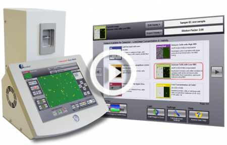
Cellometer Auto 2000 Cell Viability Counter
Touchscreen fluorescent automated cell counter for primary samples
“…far superior to any other method…”
“… quick, reliable, easy to use …”
“It is an awesome cell counter.”
“This machine [Cellometer Auto 2000] works great!”
Introduction to the Cellometer Auto 2000 Cell Viability Counter
Simple, User-friendly Procedure
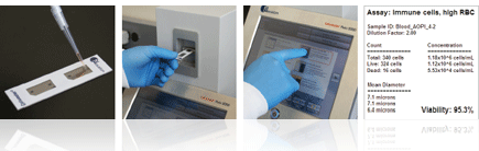
- Pipette 20 µl
- Insert Slide
- Select Assay & Click Count
- View Results in 30 seconds!
Simple, Automated Cell Counting in 30 Seconds
The Auto 2000 utilizes bright field imaging and dual-fluorescence imaging to quickly and accurately identify and count individual cells. Cell count, concentration, diameter, and % viability are automatically calculated and reported.
Load Sample, View Image, Count Cells, and Obtain Results in less than 30 seconds
The Auto 2000 Allows Users to:
- Increase throughput
- Increase accuracy
- Improve consistency
- Ensure all data is correctly captured
- Count difficult cells (clumpy, irregular-shaped)
- Eliminate judgment errors, miscounts, interference from red blood cells and user-to-user variability
Primary Cell Analysis: PBMCs, Stem Cells, and more
The Cellometer Auto 2000 is specifically optimized for analysis of primary cells from peripheral blood, cord blood, bone marrow, and other complex samples for use in a wide range of research areas, including:
- Nucleated Cells for Transplantation
- PBMCs for Immunology
- Splenocytes for Vaccine Development
- Stem Cells for Cellular Therapy
- Tumor Cell Suspensions for Oncology
Dual-color fluorescence allows for staining of live and dead nucleated cells, generating accurate viability results even in the presence of debris, platelets, and red blood cells. Accurate analysis of both ‘messy’ and ‘clean’ samples enables the Auto 2000 to evaluate samples at a variety of points throughout sample processing – from initial collection to separation, to cryopreservation.
The Cellometer Auto 2000 features one-touch assays for analysis of a wide range of primary samples, including:
Nucleated Cells from Whole Blood
![]()
Stem Cells
![]()
PBMCs / Splenocytes following Separation
![]()
Tumor and Tissue Cell Suspension
![]()
No Interference from Red Blood Cells, Platelets, or Debris
The dual-fluorescence AO/PI method utilizes nuclear staining dyes that bind to nucleic acids in the cell nucleus. Because most mature mammalian red blood cells do not contain nuclei, only live and dead mononuclear cells produce a fluorescent signal. There is no need to lyse red blood cells, saving time and eliminating an extra sample preparation step. Red blood cells, platelets, and debris are not counted in the fluorescent channels.
The advantage of fluorescent counting for primary cells
These images (right) demonstrate the advantage of fluorescent counting for primary cells. The bright field image shows the combination of nucleated cells, red blood cells, and platelets present in the sample. Only the live and dead nucleated cells are visualized and counted in the green and red fluorescent channels.
| Sample | Measurement | Total nucleated | All | RBC | % RBC | n |
|---|---|---|---|---|---|---|
| PBMC+RBC | Mean
CV |
1.26E+07
6.2% |
1.39E+07
8.8% |
1.23E+06 | 8.9% | 10 |
| PBMC+1/2RBC | Mean
CV |
1.21E+07
4.8% |
1.27E+07
5.4% |
5.82E+05 | 4.6% | 10 |
| PBMC+1/4RBC | Mean
CV |
1.22E+07
7.9% |
1.24E+07
7.3% |
2.27E+05 | 1.8% | 10 |
Fresh human PBMCs (peripheral blood mononuclear cells) were spiked with varying amounts of RBCs (red blood cells.) All cells (nucleated + RBC) were counted in the bright field channel. Nucleated cells were then counted in the green fluorescent channel. Varying amounts of RBCs (1.8%, 4.6%, and 8.9%) did not affect the nucleated cell count.

Several red blood cells are indicated in the bright field image (above, left). The red blood cells are not visible in the fluorescent image (above, right) detecting cells stained with nuclear staining dye.
Analysis of Clumpy and Irregular-shaped Cells
Including NCI-60 and clumpy MCF-7 Cells
NCI-60 is a group of 59 human cancer cell lines (originally 60) developed by the National Cancer Institute for screening purposes.
- 57% of the NCI-60 cell lines are clumpy, contain debris, or display large variations in cell shape or size
- All 59 NCI-60 cell lines have been successfully validated on the Cellometer Cell Counter
All 40 of the NCI Comprehensive Cancer Centers use Cellometer Cell Counters.

Clumpy Cells
The MCF-7 breast cancer cell line can be very clumpy. The Cellometer pattern-recognition software identifies and counts individual cells within these cell clumps for accurate analysis (shown above).

Irregular-shaped Cells
The Cellometer cell roundness setting can be adjusted for recognition and counting of irregular-shaped cells, such as RD cells and activated T-cells.
User-Friendly Touch Screen

The user-friendly touch screen displays pre-optimized assays for common cell types. User-specific protocols can be easily created and saved to the menu. The simple, pre-optimized assays make the instrument easy to operate and simplify training for new users.
With the click of a button, researchers can select a new assay with saved counting parameters, making the instrument ideal for labs testing a variety of cell types. Auto-save options allow users to quickly test a series of samples.
Live / Dead Nucleated Cell Counts using Dual-Fluorescence
Green fluorescent live cell image

Red fluorescent dead cell image

Why Dual-Fluorescence?
Because bright field cell counting does not differentiate nucleated from non-nucleated cells and trypan blue staining is not as easy to detect as fluorescent staining, dual-color fluorescence is strongly recommended for accurate viability analysis for primary cells. The Auto 2000 is equipped with standard assays for dual-fluorescence analysis of primary cells stained with AO/PI.
The AO/PI Method
Acridine Orange, AO, is a nuclear staining (nucleic acid binding) dye permeable to both live and dead cells. It stains all nucleated cells to generate green fluorescence. Propidium iodide, PI, can only enter dead cells with compromised membranes. It stains all dead nucleated cells to generate red fluorescence. Cells stained with both AO and PI fluoresce red due to quenching, so all live nucleated cells fluoresce green and all dead nucleated cells fluoresce red.
Cell Images for Data Verification
No two cells are the same.
With the Auto 2000 Cell Counter, cell morphology can be immediately viewed on-screen in the bright field image.
Counted cells are indicated on-screen for further verification that cells in the sample are being imaged and analyzed properly. Bright field counted images can be viewed for basic cell counting and trypan blue viability.
Fluorescent counted images indicating counted live and dead nucleated cells can be viewed for dual-fluorescence primary cell viability assays.
Users can confirm that:
- cells are counted correctly, based on size and shape
- cells within clumps are being counted individually
- red blood cells, platelets, and debris are being excluded from results


The bright field image confirms that individual cells within pairs are being counted and smaller debris is not being counted. In the combined fluorescent counted image, live counted cells are circled in green. Dead counted cells are circled in red.
- Cell images can be archived and exported for use in publications and presentations.
- Saved images can be re-counted using default or user-optimized analysis settings
Cell Size Analysis & Size-based Counting

The Auto 2000 Cell Counter Automatically generates a cell size histogram based on cell diameter.
Because Cellometer generates individual cell size measurements, multiple samples can be overlaid on one histogram enabling analysis of the change in cell diameter over time

For experiments involving SF9 or SF21 cells, saved images can be viewed and re-counted to analyze changes in morphology over time.

The minimum and maximum cell diameter settings can be optimized to count specific cells in a sample.
This example demonstrates counting of mature dendritic cells cultured from PBMCs based on cell diameter.
10x Faster than Manual Counting
Counting 1 x 10⁶ cells takes approximately 5 minutes with a manual hemacytometer. Counting live and dead cells sometimes takes twice as long. The Cellometer Auto 2000 Cell Viability Counter calculates cell count and concentration for live and dead cells and % viability in just 30 seconds.

Improve Data Accuracy & Consistency
- Eliminate Wash Steps
- Eliminate Judgment Errors
- Eliminate interference from RBCs
- Eliminate Recording & Calculation Errors
- Reduce Counting Time … Run More Experiments
Cellometer Precision
The Cellometer Auto 2000 offers excellent reproducibility, with a %CV (Coefficient of Variation) of <10% for fluorescent concentration and viability measurements. The data (below) is based on four preparations of Jurkat cells stained with propidium iodide, a fluorescent nuclear-staining dye.
| Sample | N Value | Average Live Cell Concentration | % Viability | CV of Concentration | CV of Viability |
|---|---|---|---|---|---|
| A | 4 | 4.20E+06 | 91.1 | 10% | 2% |
| B | 4 | 1.06E+06 | 22.7 | 7% | 1% |
| C | 4 | 3.27E+06 | 57.5 | 7% | 7% |
Imaging / Counting Chambers: No Washing or Contamination

The disposable Cellometer Cell Counting Chambers offer several key advantages:
Time savings – no washing
No risk of cross-contamination
Reduced biohazard risk to users
Controlled sample volume
Large-depth chambers for large cells
Most affordable automated counting consumables
Auto-Save Data and Cell Images

The Auto 2000 allows users to generate specific folders to which data reports and images can be auto-saved. Using the auto-save feature ensures all data is accurately captured in the correct location. It also enables users to test a series of samples very quickly

Users can configure the Auto 2000 to request a new sample name for each sample that is imaged and counted or apply auto-numbering to individual samples in a series to further reduce analysis time. Users can also apply a time stamp to individual data files.
Dedicated On-line and On-site Applications Support
Experienced Nexcelom Technical Support Specialists are available from 8:30am to 5:00pm EST for phone and on-line support and can assist with:
- Creation of new cell types
- Optimization of counting parameters
- Troubleshooting
- Training of new users
- Installation of the Auto 2000 Cell Viability Counter

The help button at the bottom right of the Cellometer Auto 2000 software screen gives users instant access to:
- Software features and instructions
- On-line tutorials and training videos
- Submission of a Support Ticket
All Nexcelom Applications Specialists are 100% focused on image-based cell concentration & viability and cell-based assays using Cellometer Image Cytometry.
Nexcelom Field-based Applications Specialists are also available for:
- On-site demonstrations
- Training
- Troubleshooting
- Technical Seminars
Applications for Cellometer Auto 2000 Cell Viability Counter

Adipocytes
Automatically measure cell size of freshly isolated adipocytes and plot size histogram. DNA staining fluorescence dyes are used to identify cells from lipid droplets. »

Adoptive Cell Transfer Therapy
Use Cellometer to perform cell based assays and measure cell size, viability and concentration of cell lines and primary samples used in adoptive cell therapy research. »

Cell Viability Measurement Using Trypan Blue or AO/PI
When should you use trypan blue and when should you use acridine orange/propidium iodide to measure cell viability? »

Cell Viability and Necrosis
Cell viability is performed using various fluorescent membrane exclusion dyes, such as PI, EB, 7AAD, and others. This assay is performed by enumerating cells in captured bright-field and fluorescent images. And Necrotic cells are detected using propidium iodide. »

TNC Concentration & Viability for Clinical (Blood) Samples
Analyze fresh and processed blood and bone marrow samples without lysing: no interference from RBCs. »

Cell Size Assay
Performing cell size measurement assay and using cell size to count cells within preset cell size parameters. For adipocytes, stem cells, Sf9 cells, dendritic cells, and others. »

NCI-60 Cancer Cell Lines
Automatically measure live cell concentration and viability of cancer cell lines used in oncology research and most of all biology research. »

Quantitative Measurement of GFP Transfection
Rapidly identify fluorescence positive cells from a sample, analyze individual cell fluorescence intensity, calculate cell concentration, size and determine the GFP transfection automatically. »

Immunology Research
Automatically quantify cell viability and concentration for a variety of immunologically relevant samples such as: bone marrow, cord blood, slpenocytes, lymphocytes, isolated mononuclear cells, tumor digests, murine samples, and others. »

Insect Cells
Automatically measure live cell concentration, viability for baculovirus infected insect cells. Cell size histogram live cell concentration and viability are generated within less than 60 seconds using 20 µl sample. »

Peripheral Blood Mononuclear Cells (PBMC)
Automatically measure live cell concentration and viability without lysing red blood cells for consistent results from patient samples. Other cells include splenocytes and bone marrow. »

Platelets
Automatically measure mouse and human platelet concentration using brightfield only method. »

Stem Cells
Automatically measure live cell concentration and viability for stem cells, such as human and mouse ES cells, mesenchymal, cardiac, induced pluripotent stem cells. Multiple viability stains, such as trypan blue, propidium iodide are used to identify dead cells. For samples with a lot of debris, dual fluorescent nuclear stains are used for live and dead cells. »

WBCs in Whole Blood
Automatically measure nucleated cell concentration without lysing red blood cells using nuclear staining dyes (AO), for human and mouse blood. »
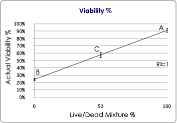
Stem Cells, Primary Cells, Cell Line
The results indicate the accuracy of the Cellometer Auto 2000 instrument in assessing the viability of Jurkat cells using AO/PI for cell viability. Four measurements were performed for each sample. The viability average was calculated and plotted. The results show the reliability and accuracy of the Cellometer Auto 2000 in measuring cell concentration and viability of mammalian cells.
 Figure 2: Chart of the % viable cells at 3 different live/dead mixture ratios. N=4 per measurement. The error bars correspond to the standard deviation for each measurement.
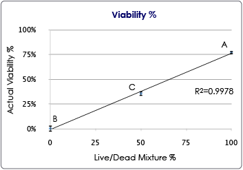 Figure 2: Chart of the % viable cells at 3 different live/dead mixture ratios. N=4 per measurement. The error bars correspond to the standard deviation for each measurement.
| Sample | N Value | Average Live Cell Concentration | % Viability | CV of Concentration | CV of Viability |
|---|---|---|---|---|---|
| A | 4 | 4.10E+06 | 78 | 1% | 2% |
| B | 4 | 0.0025E+06 | 0.6 | N/A | N/A |
| C | 4 | 2.48E+06 | 36 | 4% | 7% |
Figure 1: Table of results for AO/PI viability using the Cellometer Auto 2000.
Cell Line Viability using Propidium Iodide Only
The results indicate the accuracy of the Cellometer Auto 2000 instrument in assessing the viability of Jurkat cells using PI for cell viability. Four measurements were performed for each sample. The viability average was calculated and plotted. The results show the reliability and accuracy of the Cellometer Auto 2000 in measuring cell concentration and viability of mammalian cells.

Figure 2: Chart of the % viable cells at 3 different live/dead mixture ratios. N=4 per measurement. The error bars correspond to the standard deviation for each measurement.
| Sample | N Value | Average Live Cell Concentration | % Viability | CV of Concentration | CV of Viability |
|---|---|---|---|---|---|
| A | 4 | 4.20E+06 | 91.1 | 10% | 2% |
| B | 4 | 1.06E+06 | 22.7 | 7% | 1% |
| C | 4 | 3.27E+06 | 57.5 | 7% | 7% |
Figure 1: Table of results for cell viability using PI only.
Cell Line Total Cell Concentration
The results indicate the accuracy of the Cellometer Auto 2000 instrument cell concentration. Serial dilutions (2-, 4-, and 8-fold) were prepared using PBS and Jurkat cells. Four measurements were performed for each sample and the averages were calculated and plotted. A linear correlation between the concentration measurements and 1/dilution factor was observed. The results show the reliability and accuracy of the Cellometer Auto 2000 in measuring cell concentration.
Figure 2: Jurkat cell concentrations at various sample dilution factors. N=4. Error bars correspond to the Standard Deviations for cell concentration.
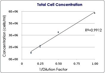
| Sample | Dilution Factor | Average Cell Concentration | CV of Concentration | Standard Deviation | N Value |
|---|---|---|---|---|---|
| A | 1 | 5.80E+06 | 1% | 4.8E+04 | 4 |
| B | 2 | 3.39E+06 | 2% | 8.12E+04 | 4 |
| C | 4 | 1.63E+06 | 6% | 9.54E+04 | 4 |
| D | 8 | 8.27E+05 | 6% | 5.33E+04 | 4 |
Figure 1: Table of results for total cell concentration for the Cellometer Auto 2000 instrument.
Low Concentration of Cells using Acridine Orange
The results indicate the accuracy of the Cellometer Auto 2000 instrument when counting Jurkat cells at very low concentrations (between 5-7E+04 cells/ml). AO was used to measure the concentration of cells in the F1 channel. Results from 4 measurements were plotted. The CV values are very close, which indicates the consistency of the measurements. The results show the reliability and accuracy of the Cellometer Auto 2000 in measuring at low cell concentration.
Figure 2: Graph of Concentration for 4 different measurements using the Low Concentration of Cells Assay and AO on the Cellometer Auto 2000.
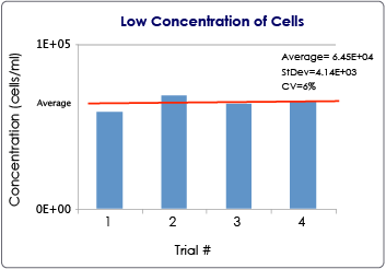
| Low Concentration AO | F1 Concentration |
|---|---|
| Sample 1 | 5.91E+04 |
| Sample 2 | 6.40E+04 |
| Sample 3 | 6.58E+04 |
| Sample 4 | 6.90E+04 |
| Average | 6.45E+04 |
| STDEV | 4.14E+03 |
| CV | 6% |
Figure 1: Table of results from the Low Concentration of Cells assay.
Performance of the Cellometer Auto 2000 Cell Viability Counter
Consistency and Accuracy Comparison to Hemacytometer
Jurkat
| N = 20 | Hemacytometer | Cellometer Auto 2000 |
|---|---|---|
| Average | 1.03E+06 | 1.01E+06 |
| STDEV | 6.60E+04 | 8.87E+04 |
| %CV | 6.4% | 8.8% |
5 µm beads
| N = 20 | Hemacytometer | Cellometer Auto 2000 |
|---|---|---|
| Average | 1.07E+06 | 1.03E+06 |
| STDEV | 5.90E+04 | 4.30E+04 |
| %CV | 5.5% | 4.2% |
Specifications for Cellometer Auto 2000 Cell Viability Counter
| Cellometer Auto 2000 | |
|---|---|
| Includes | • Cellometer Auto 2000 Instrument with integrated computer and touch-screen and dual-fluorescent optics • Cellometer Software • Two USB 2.0 ports • Power Supply • Ethernet port • Phone/online applications support during set-up |
| Imaging Performance | Cell Size: 5 – 300* microns Conc. Range: 105 – 107 cells/mlBright field imaging, fluorescent imaging and pattern-recognition software to quickly and accurately decluster, identify and count individual cells. *Cellometer CHT4-PD300 Slides are required for cells > 80 microns in diameter |
| Instrument Specifications | Weight: 26 lbs. (11.8 kg) Dimensions: Width: 11.1″ (28.3 cm) Depth: 12.8″ (32.4 cm) Height: 13.4″ (34.0 cm)Input to Power Adapter: 100-240 VAC, 50/60 Hz, 1.5A Output to Instrument: 12 VDC, 7.5A |
| Display | 10.4″ touch screen |
| Fluorescent Optics | Excitation / Emission: 470 nm/535 nm Example Fluorophores: • Acridine Orange (+DNA) • Calcein AM • CFDA • SYTO® 9 • SYTO® 13Excitation / Emission: 540 nm/605 nm Example Fluorophores: • Propidium Iodide • Ethidium Bromide |
Resources
Manuals
Brochure
Videos
- How to Improve your Primary Cell Analysis: Make it Accurate, Quick and Simple
- How to Measure Primary Cell Concentration & Viability using Cellometer Auto 2000
- Cellometer Auto 2000 Assay Overview Video
- Cellometer Auto 2000 Video Demonstration
Application Notes
- Enumeration and Viability of Nucleated Cells from Bone Marrow, Cord Blood, and Mobilized Peripheral Blood
- Concentration & Viability of PBMCs without Lysing
- Recommended procedure for comparison of cell concentration and viability using the Cellometer Auto 2000 automated cell viability counter and a manual hemacytometer
Training Webinars
- Optimizing Fluorescent Assay Parameters on the Cellometer Auto 2000
- How to Optimize Bright Field Parameters to Exclude Debris using Cellometer Auto 2000
- How to create, edit and adjust fluorescent parameters for your assays on the Cellometer Auto 2000
- Optimizing your Exposure Time and Focus Settings on the Cellometer Auto 2000
Training Videos

Getting Started with the Auto 2000
Customer Testimonials
Saved us a lot of valuable time
The Auto 2000 has improved our counting accuracy and consistency but more importantly has saved us a lot of valuable time that we now can dedicate to other work. We love the Auto 2000!
function positionLinkBlock(targetContainer) { if (targetContainer != null) { var strLinkBlock = 'UCLA
You can trust your results
I enjoy the ease of use of the Auto 2000 – everyone in the lab uses it and we have consistent results when counting samples. It is also helpful to see which cells are counted as dead and alive, so you can trust your results.
function positionLinkBlock(targetContainer) { if (targetContainer != null) { var strLinkBlock = 'Visualize
The AO/PI live/dead dye used with the Cellometer Auto 2000 is a great way to see and measure the proportion of live and dead cells in suspension. You can visualize if the dead cells are dispersed evenly or if clumping is a factor in cell viability.
function positionLinkBlock(targetContainer) { if (targetContainer != null) { var strLinkBlock = 'Very convenient
This instrument [Cellometer Auto 2000] is very convenient especially for counting the cell number of multiple samples. However, I find that it is a bit difficult to uncover the cover membrane of the slide.
function positionLinkBlock(targetContainer) { if (targetContainer != null) { var strLinkBlock = 'The Jackson laboratory
Very satisfied
Using the Auto 2000 has saved us a lot of time that would have been wasted on using traditional counting methods. The counter has proven to be very reliable when doing counts in duplicates. Overall, we are very satisfied with this equipment.
function positionLinkBlock(targetContainer) { if (targetContainer != null) { var strLinkBlock = 'A vast improvement
We are in the process of validating the instrument [Cellometer Auto 2000] prior to going live. However, the instrument is very easy to use and is a vast improvement over our previous manual counting method.
function positionLinkBlock(targetContainer) { if (targetContainer != null) { var strLinkBlock = 'Donald Hudspeth
Very important in our daily tasks
We use the Cellometer [Auto 2000] every day here in our lab. It is very important in our daily tasks, as well as contributing to our research studies. It is very straightforward to use.
function positionLinkBlock(targetContainer) { if (targetContainer != null) { var strLinkBlock = 'Far superior to any other method
We love our Auto 2000 cell counter. It is so much more accurate than the trypan blue staining we used to do to count our cells. We count everything from CHO-k1, PMBCs, and mast cells using this machine. We particularly find this method of counting cells is far superior to any other method we have tried when counting cells from a tissue digest. Would highly recommend this machine.
Quick, reliable, easy to use
We find the Auto 2000 Cellometer to be a quick, reliable, easy to use, and reasonably affordable option for the precision being offered. This instrument is a great addition to any lab!
function positionLinkBlock(targetContainer) { if (targetContainer != null) { var strLinkBlock = 'Brett Haines
Saves tons of time on a daily basis
We really enjoy the convenience of our Nexcelom Cellometer [Auto 2000]. It saves tons of time on a daily basis. We mainly use it to count live or dead cells in a population and to maintain viable levels of NK92mi cells in the desired concentration range.
function positionLinkBlock(targetContainer) { if (targetContainer != null) { var strLinkBlock = 'An awesome cell counter
I use the Auto 2000. It is an awesome cell counter. Very easy to use and fast. My cell counts are more accurate with the Cellometer compared to other cell counters. I also found the webinars and training/support very accessible and helpful.
function positionLinkBlock(targetContainer) { if (targetContainer != null) { var strLinkBlock = 'Elstar Therapeutics
Very accurate, reliable, and easy to use
The Cellometer Auto 2000 has completely replaced the Vi-Cell and Countess in my workflow. I primarily use it for counting and checking the PBMCviability of primary cells and PBMCs using the AO/PI function. I have found it to be very accurate, reliable, and easy to use.
function positionLinkBlock(targetContainer) { if (targetContainer != null) { var strLinkBlock = 'Very accurate, reliable, and easy to use
The Cellometer Auto 2000 has completely replaced the Vi-Cell and Countess in my workflow. I primarily use it for counting and checking the PBMCviability of primary cells and PBMCs using the AO/PI function. I have found it to be very accurate, reliable, and easy to use.
It plays a crucial role
It [the Cellometer Auto 2000] is very useful and accurate. I process samples every day and it plays a crucial role. It is very easy to use and understand. I came in knowing nothing, but mastering how to use the equipment was quite easy.
function positionLinkBlock(targetContainer) { if (targetContainer != null) { var strLinkBlock = 'Comparable to hemacytometer
I like the cell images with AO/PI dye. When a cell line didn’t have GFP protein just looking at the images I could tell that there was something wrong with it. I have compared it [Cellometer Auto 2000] with a hemacytometer and they are comparable while Vi-Cell was not.
function positionLinkBlock(targetContainer) { if (targetContainer != null) { var strLinkBlock = 'Susan Wilson
Comparable to hemacytometer
I like the cell images with AO/PI dye. When a cell line didn’t have GFP protein just looking at the images I could tell that there was something wrong with it. I have compared it [Cellometer Auto 2000] with a hemacytometer and they are comparable while Vi-Cell was not.
Susan Wilson
Faster, easier, and more effective
We have been counting PBMCs on a coulter counter, and once that broke, a manual hemocytometer. The Auto 2000 is faster, easier, and more effective than either method, to say nothing of the reduced hassle of cleaning a hemocytometer or constantly flushing and priming an aperture.
function positionLinkBlock(targetContainer) { if (targetContainer != null) { var strLinkBlock = 'Harry Hurley
Faster, easier, and more effective
We have been counting PBMCs on a coulter counter, and once that broke, a manual hemocytometer. The Auto 2000 is faster, easier, and more effective than either method, to say nothing of the reduced hassle of cleaning a hemocytometer or constantly flushing and priming an aperture.
Harry Hurley
Big part of our research projects
We have been using the Nexcelom [Cellometer] Auto 2000 in our lab and we greatly enjoy using it! We have not encountered any problems with it. The instrument is easy to use and is a big part of our research projects.
function positionLinkBlock(targetContainer) { if (targetContainer != null) { var strLinkBlock = 'Extremely user-friendly
The Cellometer [Auto 2000] is extremely user-friendly, even to those who have never used one. Cell counts are consistent and images are clear.
function positionLinkBlock(targetContainer) { if (targetContainer != null) { var strLinkBlock = 'Very amenable to customization
We have been very happy with the performance of the Cellometer Auto 2000 as well as the support we got when evaluating the instrument and setting it up. Since it is easy to view what is being counted and adjust the settings, we think it is very amenable to customization and this was important for our application. The instrument is still simple to use despite the ability to adapt all the settings. We haven’t been using it very long, but haven’t had any issues with it thus far.
function positionLinkBlock(targetContainer) { if (targetContainer != null) { var strLinkBlock = 'Lindsay Sulzer
Very amenable to customization
We have been very happy with the performance of the Cellometer Auto 2000 as well as the support we got when evaluating the instrument and setting it up. Since it is easy to view what is being counted and adjust the settings, we think it is very amenable to customization and this was important for our application. The instrument is still simple to use despite the ability to adapt all the settings. We haven’t been using it very long, but haven’t had any issues with it thus far.
Lindsay Sulzer
See my cell suspension prior to counting
I really love that I can use the Cellometer Auto 2000 to see my cell suspension prior to counting. If my cells look too dense and clumpy, I can further dilute my cell suspension and test another sample to see that they are suspended in sufficient volume to get accurate counts.
function positionLinkBlock(targetContainer) { if (targetContainer != null) { var strLinkBlock = 'See my cell suspension prior to counting
I really love that I can use the Cellometer Auto 2000 to see my cell suspension prior to counting. If my cells look too dense and clumpy, I can further dilute my cell suspension and test another sample to see that they are suspended in sufficient volume to get accurate counts.
Everyone in the lab prefers the Cellometer
We have multiple counting instruments (flow cytometer, trypan blue, Scepter, Nucleocounter) but everyone in the lab prefers the Cellometer [Auto 2000] due to its reliability and ease of use.
function positionLinkBlock(targetContainer) { if (targetContainer != null) { var strLinkBlock = 'This machine works great!
This machine [Cellometer Auto 2000] works great! So much easier and more accurate than counting manually.
function positionLinkBlock(targetContainer) { if (targetContainer != null) { var strLinkBlock = 'Very quick and efficient
From what I’ve heard from my mentors, cell counting sounded like a drag: grids, manual counting, yadda yadda yadda. But with this device [Cellometer Auto 2000], cell counting is very quick and efficient. The setup work in the tube is quite simple, and counting is as easy as literally pressing a button.
Extremely useful
The Auto 2000 is extremely useful for our laboratory. We use one a lot of times during our day. Cellometer helps do experiments much faster than before.
function positionLinkBlock(targetContainer) { if (targetContainer != null) { var strLinkBlock = 'Svetlana Gapon
We are confident in the accuracy
This instrument is very helpful in our lab – we use it for AO/PI, trypan, and bright field staining for at least 5 different cell types and we are confident in the accuracy of the [Cellometer] Auto 2000.
function positionLinkBlock(targetContainer) { if (targetContainer != null) { var strLinkBlock = 'Very supportive staff
Ease of use is probably the most dominating feature [of the Cellometer Auto 2000]. Backed by a very supportive staff, using instruments or services through Nexcelom has been a pleasure.
function positionLinkBlock(targetContainer) { if (targetContainer != null) { var strLinkBlock = 'Edgard Raymond
Ease of use
Ease of use is probably the most dominating feature. Backed by a very supportive staff, using instruments or services through Nexcelom has been a pleasure.
function positionLinkBlock(targetContainer) { if (targetContainer != null) { var strLinkBlock = 'John Pleau
Customer Publications using Cellometer Auto 2000
Our customers include:
