
Apoptosis Reagents
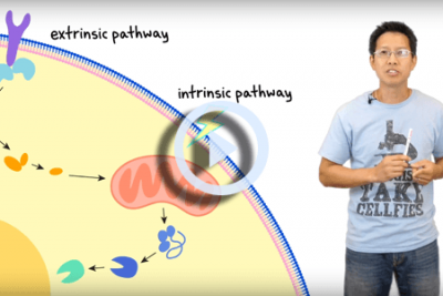
Apoptosis, or programmed cell death, is a natural process of cellular self-destruction. Apoptosis is necessary for routine cell turnover and tissue homeostasis. It is also important in embryogenesis, maintenance of immune tolerance, and development of the nervous system. Problems with the regulation of apoptosis are thought to be linked to many cancers, degenerative diseases, and autoimmune diseases, making apoptosis a key target in many fields of clinical research.
Learn more about Apoptosis »
Annexin V-FITC / PI Apoptosis Reagents (for K2, Vision CBA, Spectrum)


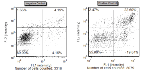
Figure 1. Scatter plots following Cellometer Vision CBA Analysis show an increase in the apoptotic cell population from 4% to 22% following incubation with 10µm α-Tos, an apoptosis-inducing drug.
Annexin V is a member of the annexin family of intracellular proteins that binds to phosphatidylserine (PS). PS is normally only found on the intracellular leaflet of the plasma membrane in healthy cells, but during apoptosis, PS translocates to the external leaflet. Fluorochrome-labeled Annexin V can then be used to specifically target and bind the PS on the surface of apoptotic cells. Cells undergoing necrosis following tissue damage or disease have compromised membranes that will also allow binding of PS. An additional dye is utilized to help differentiate necrotic cells within the population.
Propidium Iodide (PI) is a membrane-exclusion dye that permeates cells with compromised cell membranes and binds to DNA. Early apoptotic and healthy cells with intact membranes will exclude PI, while very late stage apoptotic and necrotic cells with compromised membranes are stained. Use of both Annexin V-FITC and PI allows researchers to characterize a cell population based on % normal, % apoptotic, and % necrotic /very late-stage apoptotic cells.
| Description | Catalog Number | Unit Size | Manual | SDS | |
|---|---|---|---|---|---|
| Annexin V staining solution for the detection of apoptotic cells | CS1-0114-1 | 500 uL | pdf / EU pdf | Buy Online | |
| Binding Buffer necessary for the Annexin V staining | CS0-0115-1 | 10 mL | pdf / EU pdf | Buy Online | |
| Propidium iodide for the detection of dead and/or necrotic cells | CS1-0116-1 | 500 uL | pdf / EU pdf | Buy Online | |
| Annexin V-FITC Kit is designed to detect annexin V positive cells as well as dead/necrotic propidium iodide positive cells | CSK-0117-1 | Kit | See each SDS kit components above | Buy Online |
ViaStain™ No-Wash Annexin V-FITC Kit (for Celigo)

From left to right – Bright field only. Bright field overlaid with Annexin-V, PI and Hoechst. Annexin-V, PI and Hoechst overlay.
Watch this webinar on how to run the Apoptosis assay with the Annexin V-FITC kit on the Celigo Image Cytometer.
When performing a no wash annexin-V assay in a plate, there are several challenges that we must overcome in order to achieve reliable results. Because cells are not trypsinized or washed, they remain in their natural state. This means that cells may be flat, hard to see, and hard to analyze. For this reason we counter stain the cell with Hoechst while simultaneously staining for Annexin-V and Propidium iodide (PI). This strategy allows us to easily and accurately detect, analyze, and report the percent of cells that Annexin-V and PI positive. The No-wash Annexin V-FITC Kit for Celigo includes the following features:
- A no-wash, stain and read assay for adherent cell culture. Perform your entire assay in the well of your plate (No need to trypsinize your cells)
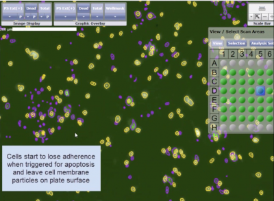
- Only nucleated cells are counted and analyzed. Cellular debris is excluded from cell population analysis
- Predefined staining, imaging, and analysis protocols are provided with the kit to allow for easy execution of the assay.
The goal of a good apoptosis assay is to analyze the cells without the need to disturb them. The no-wash Annexin-V FITC kit combined with the Celigo instrument provides that ability for both suspension and adherent cell culture.
| Description | Catalog Number | Unit Size | Manual | SDS | Buy Online |
|---|---|---|---|---|---|
| ViaStain No-Wash Annexin V-FITC Kit for Celigo | CSK-V0007-1 | Annexin V-FITC: 100 ul PI: 30 ul Hoechst: 6 ul Buffer: 10 ml |
US:Annexin V EU:Annexin V US:PI EU:PI US:Hoechst EU:Hoechst US:Buffer EU:Buffer |
Buy Online |
ViaStain™ Live Caspase 3/7 Detection for 2D/3D Culture with and without Hoechst (for Celigo)

ViaStain™ Live Caspase 3/7 Detection for 2D/3D Culture kit is designed to detect caspase 3/7 activity in live cells. The ability to perform kinetic apoptosis assays allows researchers to continuously measure the caspase 3/7 activity within the cell population. This no wash assay has been shown to effectively work in both 2D and 3D cultures.
The reagent (NucViewTM) consists of a nucleic acid-binding dye with a fluorescent probe that is attached to a four-amino acid peptide sequence DEVD (Asp-Glu-Val-Asp) forming a cell membrane-permeable DEVD-DNA complex. While the nucleic-acid dye is linked to the DEVD peptide sequence, the dye is unable to bind to DNA and remains non-fluorescent. During apoptosis, caspase 3/7 proteins cleave the DEVD-DNA dye complex and thereby release the high-affinity DNA dye, which translocates to the nucleus and binds to the DNA, producing a bright green fluorescent signal.
To enumerate the total number of nucleated cells and obtain percent of apoptotic cells, the cells can be counterstained with Hoechst 33342.
ViaStain™ Live Caspase 3/7 Detection for 2D/3D Culture (for Celigo)
| Description | Catalog Number | Unit Size | Manual | SDS | |
|---|---|---|---|---|---|
| In real-time, measure apoptosis by detecting Caspase 3/7 in 2D live cell culture and 3D tumor spheroids. | CS1-V0002-1 | 100 uL | US:Caspase 3/7 EU:Caspase 3/7 |
Buy Online | |
| In real-time, measure apoptosis by detecting Caspase 3/7 in 2D live cell culture and 3D tumor spheroids and determine total number of cells using Hoechst. | CSK-V0003-1 | 100 uL | US:Caspase 3/7 EU:Caspase 3/7 US:Hoechst EU:Hoechst |
Buy Online |
Caspase-3 Apoptosis Kit (for Vision CBA)

Caspase-3 belongs to a family of evolutionally conserved cysteine proteases that play a key role in regulating programmed cell death, or apoptosis. Caspase-3 activation triggers a series of irreversible events that lead to the death of the cell, including activation of the CAD endonuclease that degrades DNA within the nucleus and initiates chromatin condensation. Increasing evidence suggests involvement of caspase-3 in Alzheimer’s Disease (AD) via cleavage of amyloid prescursor protein (APP) and tau and the formation of beta-amyloid (Aβ) and neurofibrillary tangles (NFTs). Other studies have demonstrated a role for caspase-3 in growth stimulation via PGE2. High amounts of caspase-3 have been linked to an increased rate of cancer recurrence and death in patients undergoing radiation treatment through the stimulation of growth of surviving cancer cells, making caspase-3 a key prognostic marker and therapeutic target in cancer therapy.

| Cell P0pulation | % of Cells | Concentration (10∧6 cells/mL) |
|---|---|---|
| Total | 100 | 2.99 |
| Caspase + | 40.16 | 1.20 |
| Caspase – | 59.84 | 1.79 |
Caspase-3 Activity histogram (left) and data table (above) for a Jurkat cell sample following overnight incubation with 10µm α-Tos, an apoptosis-inducing drug.
Principle of the Cellometer Caspase-3 Apoptosis Assay
The CaspGLOW™Fluorescein Active Caspase-3 Staining Kit offers a simple, sensitive method for detection of active Caspase-3 in living cells. When detecting caspase-3, we utilize a specific FITC-conjugated caspase-3 inhibitor, DEVD-FMK. DEVD-FMK is cell-permeable and binds irreversibly to active caspase-3 within the apoptotic cell. The fluorescent marker (FITC) on the FITC-DEVD-FMK allows for the detection of cells undergoing apoptosis via Caspase-3 activation using image cytometry. Each kit contains ready-to-use reagents sufficient for 100 tests along with a detailed instruction manual outlining cell staining, imaging, and cell cycle data analysis using the Cellometer Vision CBA Analysis System.
CaspGLOW™ Fluorescein in Active Caspase-3 Staining Kit
| Description | Catalog Number | Unit Size | Manual | MSDS | |
|---|---|---|---|---|---|
| CaspGLOW Fluorescein Active Caspase-3 Staining Kit for Apoptosis. | K183-25-N
K183-100-N |
25 uL
100 uL |
—
— |
—
— |
Buy Online |
Caspase-8 Apoptosis Kit (for Vision CBA)

The Cellometer Caspase-8 Apoptosis Kit is designed for fast, simple determination of Caspase-8 activity for a cell population using image cytometry. Each kit contains ready-to-use reagents sufficient for 100 tests along with a detailed instruction manual outlining cell staining, imaging, and cell cycle data analysis using the Cellometer Vision CBA Analysis System.

| Cell P0pulation | % of Cells | Concentration (10∧6 cells/mL) |
|---|---|---|
| Total | 100 | 3.88 |
| Caspase + | 27.54 | 1.07 |
| Caspase – | 72.17 | 2.80 |
Caspase-8 Activity histogram (left) and data table (above) for a Jurkat cell sample
Principle of the Cellometer Caspase-8 Apoptosis Assay
The CaspGLOW™Fluorescein Active Caspase-8 Staining Kit offers a simple, sensitive method for detection of active Caspase-8 in living cells. When detecting caspase-8, we utilize a specific FITC-conjugated caspase-8 inhibitor, IETD-FMK. IETD-FMK is cell-permeable and binds irreversibly to active caspase-8 within the cell. The fluorescent marker (FITC) on the IETD-FMK allows for the detection of cells undergoing apoptosis via Caspase-8 activation using the Vision CBA Analysis System. Results are presented as a Caspase positive population percentage and cell concentration.
CaspGLOW™ Fluorescein in Active Caspase-8 Staining Kit
| Description | Catalog Number | Unit Size | Manual | MSDS | |
|---|---|---|---|---|---|
| CaspGLOW Fluorescein Active Caspase-8 Staining Kit for Apoptosis. | K188-25-N
K188-100-N |
25 uL
100 uL |
—
— |
—
— |
Buy Online |
Instrument Compatibility for Apoptosis Reagents
| Auto 2000 | K2 | Vision CBA | Spectrum | Celigo | |
|---|---|---|---|---|---|
| Annexin V-FITC | X | X | X | ||
| Annexin V Binding Buffer | X | X | X | ||
| ViaStain™ PI Staining Solution for Apoptosis | X | X | X | X | |
| ViaStain No-Wash Annexin V-FITC Kit | X | ||||
| ViaStain™ Live Caspase 3/7 Detection for 2D/3D Culture | X | ||||
| ViaStain™ Live Caspase 3/7 Detection for 2D/3D Culture with Hoechst | X | ||||
| CaspGLOW™ Fluorescein in Active Caspase-3 Staining Kit | X | ||||
| CaspGLOW™ Fluorescein in Active Caspase-8 Staining Kit | X |
Cellaca PLX Apoptosis Detection Kits
| Description | Catalog Number | Unit Size | Manual | SDS |
|---|---|---|---|---|
| Cellaca® PLX, Annexin V-FITC/PI Apoptosis Kit | CSK-A0029-1 | 25 tests | ||
| CSK-A0029-2 | 100 tests | |||
| Cellaca® PLX, Caspase 3/RubyDead Apoptosis Kit | CSK-A0028-2 | 100 tests |
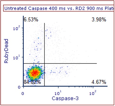
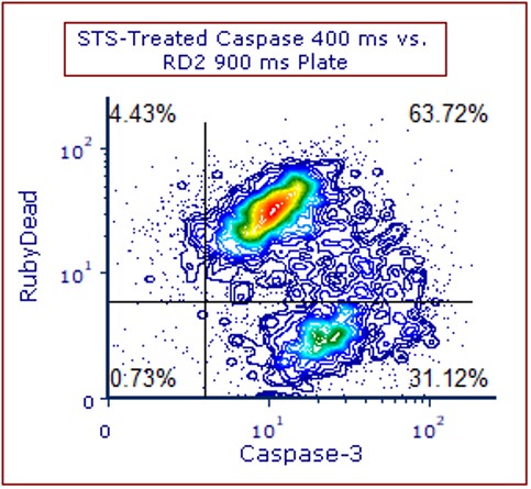
Above data is showing untreated (control) and staurosporine (drug) treated Jurkat cells stained with Caspase 3 and RubyDead dyes. In the untreated sample, healthy cells can be seen in the lower left-and quadrant. In the drug treated sample, majority of the cells are either caspase 3 positive (in lower right-hand quadrant) or caspase 3 and RubyDead positive (top right).