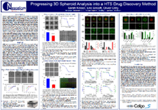
Progressing 3D Spheroid Analysis into a HTS Drug Discovery Method

This study highlights the use of the 384-well low attachment U-bottom plate combined with the Celigo® Imaging Cytometer to image and analyze the formation of 3D spheroids. This allows for increased throughput, number of replicates and parameters per plate as compared to 96-well plates. U87MG cells were used to create tumorspheres in 384-well plates and were subsequently analyzed by imaging. The data illustrate that reproducible 3D spheroids can be formed in 384-well plates.
