Adherent Cell Cycle PI with FCS Express
Presenter: Sarah Kessel, Nexcelom Applications Specialist
Assay: Cell Counting Techniques
Instrument: Celigo
Runtime: 00:50:24
Aired: March 14, 2019
How to Run an Apoptosis Assay on the Celigo Using Annexin V PI Hoechst
Presenter: Sarah Kessel, Nexcelom Applications Specialist
Assay: Cell Counting Techniques
Instrument: Celigo
Runtime: 00:37:28
Aired: November 8, 2018
How to Create and Use Project Mode for Growth Tracking with Direct Cell Counting on the Celigo
Presenter: Sarah Kessel, Nexcelom Applications Specialist
Assay: Cell Counting Techniques
Instrument: Celigo
Runtime: 00:45:33
Aired: September 17, 2018
How to Capture Bright Field Images along with Viability Images on the Celigo
Presenter: Sarah Kessel, Nexcelom Applications Specialist
Assay: Cell Counting Techniques
Instrument: Celigo
Runtime: 00:32:22
Aired: August 9, 2018
How to Prepare and Analyze and Antibody Binding Assay on the Celigo
Presenter: Sarah Kessel, Nexcelom Applications Specialist
Assay: Cell Counting Techniques
Instrument: Celigo
Runtime: 00:32:56
Aired: June 15, 2018
In this webinar:
- Assay setup
- Image acquisition
- Image analysis
- Data interpretation
How to Run a High Throughput Viability Assay on Celigo Suspension Cells using AO/PI Stain
Presenter: Sarah Kessel, Nexcelom Applications Specialist
Assay: Cell Counting Techniques
Instrument: Celigo
Runtime: 00:47:49
Aired: April 12, 2018
Nexcelom’s Applications Scientist Sarah Kessel explains quick and easy plate preparation, image acquisition & analysis, as well as exporting data into a template.
Setting up the Celigo for Successful Proliferation Experiments using Confluence Measurement
Presenter: Sarah Kessel, Nexcelom Applications Specialist
Assay: Celigo Software
Instrument: Celigo
Runtime: 00:43:53
Aired: Mar 15, 2018
How to Better Manage Your Data on Celigo
Presenter: Sarah Kessel, Nexcelom Applications Specialist
Assay: Celigo Software
Instrument: Celigo
Runtime: 00:49:00
Aired: Nov 30, 2017
Nexcelom’s Applications Scientist Sarah Kessel helps demonstrate how to import, export, and delete scans as well as how to create folders and new users with permissions.
Getting Better Acquainted with the Celigo Gating Tab
Presenter: Sarah Kessel, Nexcelom Applications Specialist
Assay: Celigo Software
Instrument: Celigo
Runtime: 00:57:41
Aired: Sep 14, 2017
Nexcelom’s Applications Scientist Sarah Kessel is bringing you a training webinar on how to become more familiar with the Gating Tab within the Celigo software.
Getting to Know the Celigo Analyze Tab Parameters Better
Presenter: Sarah Kessel, Nexcelom Applications Specialist
Assay: Celigo Software
Instrument: Celigo
Runtime: 00:37:49
Aired: Aug 10, 2017
Nexcelom’s Applications Scientist Sarah Kessel is bringing you a training webinar on how to become more familiar with the Analyze Tab within the Celigo software.
Detect Apoptosis in your cells with Caspase 3/7 and Hoechst on Celigo
Presenter: Sarah Kessel, Nexcelom Applications Specialist
Assay: Apoptosis
Instrument: Celigo
Runtime: 00:28:33
Aired: July 6, 2017
Nexcelom’s Applications Scientist Sarah Kessel is bringing you a training webinar on how to detect Apoptosis in your cells with Caspase 3/7 and Hoechst on Celigo. We will cover how to set up the assay all the way through reviewing and exporting data and images.
Perform Label-free Proliferation Experiments on Adherent Cells using Celigo
Presenter: Sarah Kessel, Applications Scientist, Nexcelom Bioscience
Assay: Proliferation
Instrument: Celigo
Runtime: 00:42:38
Aired: June 8, 2017
Nexcelom’s Applications Scientist Sarah Kessel is bringing you a training webinar on how to preform label-free proliferation experiments on adherent cells using Celigo. We will cover how to set up the assay, how to acquire the images, how to set the image analysis parameters, how to export your data and create familiar graphs.
Celigo Viability Assays with Hoechst & PI
Presenter: Sarah Kessel, Applications Scientist, Nexcelom Bioscience
Assay: Viability
Instrument: Celigo
Runtime: 00:32:33
Aired: April 27, 2017
Nexcelom’s Applications Scientist Sarah Kessel is bringing you a training webinar on how to obtain viability measurements with Hoechst & Propidium Iodide on Celigo. We will cover how to set up the assay, how to acquire the images, how to set the image analysis parameters, how to export your data and create familiar graphs.
How to Run a High-Throughput Viability Assay on Suspension Cells using AO/PI stain on your Celigo
Presenter: Sarah Kessel, Applications Scientist, Nexcelom Bioscience
Assay: Viability
Instrument: Celigo
Runtime: 00:42:05
Aired: April 13, 2017
Nexcelom’s Applications Specialist team is bringing you a training webinar on how to run a high-throughput viability assay on suspension cells, using a nuclear viability stain, Acridine Orange (AO) and Propidium Iodide (PI) on the Celigo.
How to run a High-Throughput Antibody Internalization Assay with pHrodo
Presenter: Sarah Kessel, Applications Scientist, Nexcelom Bioscience
Assay: Antibody Internalization
Instrument: Celigo
Runtime: 00:39:16
Aired: February 9, 2017
In this user training webinar, Sarah Kessel, Applications Scientist at Nexcelom, brings a how-to video on setting up and running a high-throughput antibody internalization assay using a ph-dye, such as pHrodo. Sarah starts be discussing some background information on this assay and reviews some great resources for additional information, such as reference papers that may be of use and interest. Sarah then walks viewers through the protocol, how to acquire and analyze images and then reviewing the data.
How to set up an Antibody Binding Assay on your Celigo
Presenter: Sarah Kessel, Applications Scientist, Nexcelom Bioscience
Assay: Antibody Binding
Instrument: Celigo
Runtime: 00:30:50
Aired: January 12, 2017
How to Quickly set up a 6-well plate for Monitoring your GFP-Transfected Cells on Celigo
Presenter: Sarah Kessel, Applications Scientist, Nexcelom Bioscience
Assay: Transfection Efficiency
Instrument: Celigo
Runtime: 00:43:40
Aired: December 8, 2016
In this user training webinar, Applications Scientist, Sarah Kessel, provides information on monitoring GFP transfection quickly in a 6 well plate. Sarah begins this session be reviewing technical content such as: what is the confluence ratio, how plate quality can present unique challenges and how to overcome those, what is the FOR and the FOV, what is subsampling and how can that speed up image acquisition, and what is the resolution for the Celigo, and how can adjusting the resolution speed up the assay. Sarah then walks users through the Celigo software, utilizing these factors already discussed.
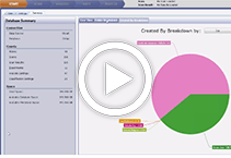
Better Organize and Manage your Data on Celigo
Presenter: Sarah Kessel, Applications Scientist, Nexcelom Bioscience
Assay: Celigo Software
Instrument: Celigo
Runtime: 00:49:37
Aired: November 11, 2016
In this user training webinar, Sarah Kessel, Applications Scientist at Nexcelom Bioscience looks at the Data Management section in the Celigo software. Sarah covers three main topics, and explores many subfeatures within each topic. She reviews the manage data page and explains: the file structure, users versus folders, filters available, and the tab options. She covers the button featues and describes how to import/export/delete and the select all or select none. Lastly, she details how to create new: users, groups and permissions. All throughout this session, Sarah also provides time saving tips and tricks.
How to Monitor 3D Multicellular Tumor Spheroid Invasion and Migration into Extracellular Matrix
Presenter: Sarah Kessel, Applications Scientist, Nexcelom Bioscience
Assay: 3D Tumor Spheroids
Instrument: Celigo
Runtime: 00:48:16
Aired: October 13, 2016
In this user training webinar, Sarah Kessel, Applications Scientist at Nexcelom Bioscience covers how to perform migration and invasion assays on 3D multicellular tumor spheroids (MCTS) on your Celigo. A big thanks is given to Steve Titus of NIH-NCATS for providing the screening compounds, and also to Mara Vinci of ICR for developing a fantastic protocol for us to use in this assay. Sarah reviews the assay protocol and the steps to set up the Celigo before moving into describing how to optimize the parameters for this experiment. Wrapping up this session, Sarah describes how to view the kinetic images that have been acquired throughout the experiment.
Getting to know the Gate Tab Parameters on Celigo
Presenter: Sarah Kessel, Applications Scientist, Nexcelom Bioscience
Assay: Celigo Software
Instrument: Celigo
Runtime: 01:03:42
Aired: August 11, 2016
In this user training webinar, Sarah Kessel, Applications Scientist at Nexcelom Bioscience reviews the various parameters in the Gate Tab in the Celigo software. Sarah presents first information about gating – what is it, why is it important, when would you use it. From there, we’ll explain everything you need to know to feel comfortable using the Gate Tab and to use it like a pro. Sarah covers: the types of plots that can be created by the Celigo – 1D Histograms, 2D Scatter Plots; the parameters that can be used to create the plots – X/Y Position, Area, Form Factor, Smoothness, Aspect Ratio, Mean or Integrated Fluorescent Intensity; the types of gates that can be drawn on the plots – Splitter, Range, Quadrant, Box, Circle, Free-Form; which Applications in Celigo utilize the Gate Tab – Data Output: Total Only, Data Putput: Total + 1 Class, Data Output: Total + 4 Classes; and how to create subpopulations and classes.
Getting to know the Analyze Tab Parameters on Celigo
Presenter: Sarah Kessel, Applications Scientist, Nexcelom Bioscience
Assay: Celigo Software
Instrument: Celigo
Runtime: 00:39:13
Aired: July 14-2016
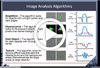
In this user training webinar, Sarah Kessel, Applications Scientist at Nexcelom Bioscience reviews the various parameters in the Analyze Tab in the Celigo software to explain the termonology and the science behind imaging and image analysis. Sarah will explain everything you need to know to feel comfortable with the buttons and settings in the Analyze Tab and to use it like a pro. Sarah covers: how the well mask localizes object identification in the well area; the different algorithms and how each work; when to adjust the precision and how it works; why “separate touching objects” works for some cell types and not others; how to eliminate objects that are not cells.
How to detect Apoptosis on Celigo: ViaStain™ Caspase 3/7 & Hoechst
Presenter: Sarah Kessel, Applications Scientist, Nexcelom Bioscience
Assay: Apoptosis
Instrument: Celigo
Runtime: 00:28:11
Aired: June 9, 2016
In this user training webinar, Sarah Kessel, Applications Scientist at Nexcelom Bioscience reviews how to perform an Apoptosis assay on adherent and suspension cells on the Celigo, using a reagent kit from Nexcelom – Nexcelom ViaStain Live Caspase 3/7 Detection for 2D/3D Culture with Hoechst (Cat# CSK-V0003-1). Sarah begins the session by reviewing the assay protocol to set up the plate before imaging on the Celigo. Sarah then covers: how to set up the assay on the Celigo – in the Start Tab ; how to acquire the images – in the Scan Tab; how to set up the image analysis – in the Analyze Tab, how to review the data and export the information and images – in the Gate and Results Tabs.
Apoptosis Annexin V Assay on Celigo
Presenter: Sarah Kessel, Applications Scientist, Nexcelom Bioscience
Assay: Apoptosis
Instrument: Celigo
Runtime: 00:46:37
Aired: May 12, 2016
In this user training webinar, Sarah Kessel, Applications Scientist at Nexcelom Bioscience reviews how to perform an Apoptosis assay on adherent cells on the Celigo, using Nexcelom reagents: Annexin V-FITC (Cat# CS1-0114 & CS0-0115-100mL), PI (Cat# CS1-0116) and Hoechst (Cat# CS1-0128-5mL). Sarah begins by explaining the concept of apoptosis and also Annexin V staining. We review the materials and reagents that will be needed, and also the assay protocol for this experiment. Sarah then covers: how to acquire and analyze images using 2 different Applications on the Celigo: PS Externalization (3 channels) and Expression Analysis (4 channels and customizable); how to use the gating tools with the Expression Analysis Application to define populations in the assay.
How to quantify phases of Cell Cycle using BrdU & DAPI stained cells imaged on Celigo
Presenter: Sarah Kessel, Applications Scientist, Nexcelom Bioscience
Assay: Cell Cycle
Instrument: Celigo
Runtime: 00:40:21
Aired: April 14, 2016
In this user training webinar, Sarah Kessel, Applications Scientist at Nexcelom Bioscience reviews how to perform an Cell Cycle assay on adherent cells on the Celigo, using BrdU and DAPI staining. Sarah begins by explaining the principle of cell cycle using BrdU (for identifying cells in synthesis) and DAPI (for nuclear staining). We review the materials and reagents that will be needed, and also the assay protocol for this experiment. Sarah then covers: how to set up the assay; how to acquire images; how to gate your cells to create the horseshoe scatter plot; how to identify to cell cycle phases; how to export your data into Excel to graph your data.
How to obtain viability measurements with Hoechst & Propidium Iodide on your Celigo
Presenter: Sarah Kessel, Applications Scientist, Nexcelom Bioscience
Assay: Viability
Instrument: Celigo
Runtime: 00:33:46
Aired: March 10, 2016
In this user training webinar, Sarah Kessel, Applications Scientist at Nexcelom Bioscience reviews how to perform a Viability assay on adherent cells on the Celigo, using Nexcelom reagents, Hoechst (Cat# CS1-0128-5mL) and PI (Cat# CS1-0109) staining. Sarah begins by reviewing the materials and reagents that will be needed, and also the assay protocol for this experiment. Sarah then covers: how to set up the assay, how to acquire the images, how to set the image analysis parameters, how to export your data and create familiar graphs.
How to analyze Wound Healing / Scratch Assay images on Celigo
Presenter: Sarah Kessel, Applications Scientist, Nexcelom Bioscience
Assay: Wound Healing
Instrument: Celigo
Runtime: 00:37:34
Aired: February 11, 2016
In this user training webinar, Sarah Kessel, Applications Scientist at Nexcelom Bioscience reviews how to perform a wound healing/scratch assay on adherent cells on the Celigo. Sarah begins by reviewing the materials and reagents that will be needed, and also the assay protocol for this experiment. Sarah then covers: how to set up the assay, how to monitor migration with label free, bright field images; how to image the wound over multiple days to produce growth curves; characterization of primary cell lines.
How to analyze Tumor Spheroids on your Celigo
Presenter: Sarah Kessel, Applications Scientist, Nexcelom Bioscience
Assay: 3D Tumor Spheroids
Instrument: Celigo
Runtime: 00:42:02
Aired: January 14, 2016
In this user training webinar, Sarah Kessel, Applications Scientist at Nexcelom Bioscience reviews how to analyze tumor spheroids on the Celigo, both label free and with viability staining using Nexcelom reagent kit, Nexcelom ViaStain(TM) Calcein AM / PI / Hoechst Cell Viability Kity (Cat# CSK-V0001-1). Sarah begins by reviewing the plate map for the experiment. Sarah then covers 2 sections. The first part reviews label free bright field imaging: setting segmentation parameters for single tumor spheroids per well; batch analysis on multiple scans; viewing wells in different scan times; exporting and graphing data. The second reviews using a fluorescent viability stain: how to use a viability stain to determine sphere health; segmenting parameters for stained spheres, exporting and graphing data.
Capture Brightfield Images along with Viability Images on Celigo
Presenter: Sarah Kessel, Applications Scientist, Nexcelom Bioscience
Assay: Viability
Instrument: Celigo
Runtime: 00:32:18
Aired: December 10, 2015
In this user training webinar, Sarah Kessel, Applications Scientist at Nexcelom Bioscience reviews how to acquire bright field images along with fluorescent, viability images of adherent cells on the Celigo using Nexcelom reagents, Hoechst (Cat# CS1-0128-5mL) and PI (Cat# CS1-0109). Sarah begins by explaining how having bright field images can be useful, to see morphological changes in your cells throughout your experiment. She then will review the materials and reagents that will be needed, and also the assay protocol for this experiment. Sarah then covers: how to acquire images in a 3-channel assay; how to identify dead cells; observing morphological changes using the bright field images; how to set up gating parameters.
Monitor GFP Transfection with Hoechst
Presenter: Sarah Kessel, Applications Scientist, Nexcelom Bioscience
Assay: Transfection Efficiency
Instrument: Celigo
Runtime: 00:21:58
Aired: August 13, 2015
In this user training webinar, Sarah Kessel, Applications Scientist at Nexcelom Bioscience reviews how to monitor GFP transfection efficiency on adherent cells on your Celigo using Nexcelom reagent, Hoechst (Cat# CS1-0128-5mL) . For this training, Sarah will cover how to acquire your images, create segmentation parameters, gate your populations and review and export your data. She breaks the training into two segments. In Part 1, she reviews how to use a single mask, to identify Hoechst-stained nuclei. In Part 2, she covers how to use a merged mask – looking at both the stained nuclei and also the GFP signal.
Monitoring of GFP Transfection Efficiency Label-free on Celigo

Presenter: Sarah Kessel, Applications Scientist, Nexcelom Bioscience
Assay: Transfection Efficiency
Instrument: Celigo
Runtime:
Aired: July 9, 2015
In this user training webinar, Sarah Kessel, Applications Scientist at Nexcelom Bioscience reviews how to measure GFP transfection efficiency on adherent cells label-free on your Celigo. For this training, Sarah breaks the training into two segments. In Part 1, she reviews how to monitor your total cell counts over time, capturing both bright field and fluorescent images. In Part 2, she addresses super confluent or difficult to identify cells, and how to measure a percentage of GFP confluence area from your total cell confluence area.
Setting up the Celigo for Successful Proliferation Experiments using Confluence Measurements

Presenter: Sarah Kessel, Applications Scientist, Nexcelom Bioscience
Assay: Proliferation
Instrument: Celigo
Runtime:
Aired: June 11, 2015
In this user training webinar, Sarah Kessel, Applications Scientist at Nexcelom Bioscience reviews how to perform proliferation experiments using confluence measurements on adherent cells on your Celigo. Sarah will review the materials and reagents that will be needed, and also the assay protocol for this experiment. Sarah then covers: how to acquire bright field images; setting up your segmentation parameters; reviewing your data; saving your experiment for additional growth tracking scans; how to load a plate with a saved experiment.
How to Perform a Viability Assay on Celigo

Presenter: Sarah Kessel, Applications Scientist, Nexcelom Bioscience
Assay: Viability
Instrument: Celigo
Runtime: 00:43:23
Aired: May 14, 2015
In this user training webinar, Sarah Kessel, Applications Scientist at Nexcelom Bioscience reviews how to perform viability experiments on adherent cells on the Celigo, with Nexcelom’s reagent kit, Nexcelom ViaStain(TM) Calcein AM / PI / Hoechst Cell Viability Kity (Cat# CSK-V0001-1). Sarah will review the materials and reagents that will be needed, and also the assay protocol for this experiment. Sarah then covers: how to set up a 3 (Red, Green & Blue) or 4 (Red, Green, Blue & Bright Field) channel assay; acquiring your images; setting up your segmentation parameters; reviewing and plotting your data.
How to Detect Cell Cycle Phases with Propidium Iodide on Celigo

Presenter: Sarah Kessel, Applications Scientist, Nexcelom Bioscience
Assay: Cell Cycle
Instrument: Celigo
Runtime:
Aired: January 8, 2015
In this user training webinar, Sarah Kessel, Applications Scientist at Nexcelom Bioscience reviews how to detect cell cycle phases on adherent cells on the Celigo, using propidium iodide staining. Sarah begins by reviewing the assay principle of cell cycle, and also the assay protocol and plate preparation for this experiment. Sarah then covers: how to acquire your images; how to set the image analysis parameters; how to export your data and create familiar graphs.
Counting Adipocytes on the Cellometer Auto 1000 and Cellometer Mini
Presenter: Lauren Bristol, Nexcelom Technical Support Specialist
Assay: Cell Counting Techniques
Instrument: Cellometer Mini,Cellometer Auto 1000
Runtime: 00:17:47
Aired: Mar 22, 2018
In this information webinar, Nexcelom’s Technical Support Specialist, Lauren Bristol helps explain which Nexcelom slides are used to count adipocytes, and the correct software settings to optimize counting.
How to Measure Percent GFP using Cellometer K2
Presenter: Lauren Bristol, Nexcelom Technical Support Specialist
Assay: Cellometer Software
Instrument: Cellometer K2
Runtime: 00:14:44
Aired: Feb 22, 2018
Learn how to use the Cellometer K2 to measure the percent GFP.
Optimizing Fluorescent Assay Parameters on the Cellometer Auto 2000
Presenter: Lauren Bristol, Technical Support Specialist
Assay: Cellometer Software
Instrument: Cellometer Auto 2000
Runtime: 00:28:17
Aired: Oct 26, 2017
Nexcelom’s Technical Support Specialist, Lauren Bristol, will cover optimization of fluorescent assay parameters on the Cellometer Auto 2000.
Bright Field vs. Fluorescence – Which Cell Imaging Method is Best for Your Cell Samples?
Presenter: Lauren Bristol, Technical Support Specialist
Assay: Cell Counting Techniques
Instrument: Cellometer-All
Runtime: 00:27:23
Aired: Sep 28, 2017
Nexcelom’s Technical Support Specialist, Lauren Bristol, will cover the differences between bright field and fluorescent imaging and which one is the best choice for your cell sample type.
Perform Accurate Trypan Blue Viability Assays on the Cellometer Auto 1000
Presenter: Lauren Bristol, Technical Support Specialist
Assay: Viability
Instrument: Cellometer Auto 1000
Runtime: 00:24:25
Aired: Aug 24, 2017
Nexcelom’s Technical Support Specialist, Lauren Bristol, will cover sample prep and the necessary steps to perform a trypan blue viability assay using Cellometer Auto 1000.
How to Perform AO/PI Viability Assays on the Cellometer K2
Presenter: Lauren Bristol, Technical Support Specialist
Assay: Viability
Instrument: Cellometer K2
Runtime: 00:18:24
Aired: July 27, 2017
Nexcelom’s Technical Support Specialist, Lauren Bristol, will cover sample prep and the necessary steps to perform an AO/PI assay on the Cellometer K2.
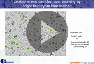
How to Count Small Cells Such as Platelets, Algae, Yeasts and Bacteria
Presenter: Dr. Leo Chan, Nexcelom Senior Scientist
Assay: Cell Counting Techniques
Instrument: Cellometer-All
Runtime: 00:20:00
Aired: July 5, 2017
Dr. Leo Chan, Nexcelom Technology R&D Manager demonstrates how to automatically count small cells using image cytometry.
How to Optimize Bright Field Parameters to Exclude Debris using Cellometer Auto 2000

Presenter: Lauren Bristol, Technical Support Specialist
Assay: Cell Counting Techniques
Instrument: Cellometer Auto 2000
Runtime: 00:31:43
Aired: June 22, 2017
Nexcelom’s Technical Support Specialist, Lauren Bristol, will cover optimization of bright field parameters on the Cellometer Auto 2000 to exclude debris from your results.
Cellometer K2 & Vision CBA Cell Cycle Analysis
Presenter: Lauren DesRoches, Technical Support Specialist, Nexcelom Bioscience
Assay: Cellometer Software
Instrument: Cellometer K2,Cellometer Vision CBA
Runtime: 00:38:59
Aired: May 25, 2017
Nexcelom’s Technical Support Specialist, Lauren DesRoches, will cover sample preparation for the cell cycle assay as well as data analysis.
Examining the most commonly asked Frequently Asked Questions on the Cellometer

Presenter: Lauren DesRoches, Technical Support Specialist, Nexcelom Bioscience
Assay: Cellometer Software
Instrument: All Cellometer
Runtime: 00:41:57
Aired: April 27, 2017
Nexcelom’s Technical Support team is bringing you a training webinar to take a look at and answer some of the most frequently asked questions they receive.
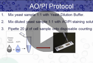
Optimizing your Yeast AO/PI Viability Assays on your Cellometer
Presenter: Lauren DesRoches, Technical Support Specialist, Nexcelom Bioscience
Assay: Yeast Viability
Instrument: Cellometer X1/X2,Cellometer Vision CBA
Runtime: 00:28:04
Aired: March 23, 2017
The Technical Support team at Nexcelom is bringing you a training webinar to walk through how to optimize your yeast AO/PI viability assays. The content covered in this webinar will be beneficial for anyone using a Cellometer X1, X2, X4, or Vision 10X instrument. Key take-aways for this webinar include: Review of the AO/PI assay principle – how it works and why we trust it. Advantages of using AO/PI over Methylene Blue for viability assays. How to use our Yeast AO/PI Viability Kit (CSK-0102). How to adjust the fluorescent parameters to ensure an accurate sample count.
Optimize your Bright Field Parameters to Exclude Debris
Presenter: Lauren DesRoches, Technical Support Specialist, Nexcelom Bioscience
Assay: Cellometer Software
Instrument: All Cellometer
Runtime: 00:25:25
Aired: February 23, 2017
Whether you are a current Cellometer user or are considering becoming a Cellometer user, we want to make sure you feel as confident and comfortable using the instrument as possible. Nexcelom’s Technical Support team is bringing you a training webinar to walk through how to optimize your bright field parameters to exclude debris when performing cell counts and viability assays. The content covered in this webinar will be beneficial for anyone using bright field imaging for count, concentration and Trypan Blue viability. The Cellometer Auto T4 software will be used for demonstration purposes, but the training can be applied to any Cellometer software.
How to create, edit and adjust fluorescent parameters for your assays on the Cellometer Auto 2000
Presenter: Lauren DesRoches, Technical Support Specialist, Nexcelom Bioscience
Assay: Cellometer Software
Instrument: Cellometer Auto 2000
Runtime: 00:30:33
Aired: January 26, 2017
In this user training webinar, Lauren DesRoches, Technical Support Specialist at Nexcelom, pulls up the Cellometer Auto 2000 software and walks you through how to create, edit and adjust the fluorescent parameters. Main areas of focus for this session are the following fluorescent settings: normalized intensity, non-uniform fluorescence, free nuclei, combined BF/FL mode, fluorescent threshold. Lauren discusses and explains what each of these parameters are and do, and then demonstrates how changing these settings can impact how the software counts the samples.
Optimizing your Exposure Time and Focus Settings on the Cellometer Auto 2000
Presenter: Lauren DesRoches, Technical Support Specialist, Nexcelom Bioscience
Assay: Cellometer Software
Instrument: Cellometer Auto 2000
Runtime: 00:31:56
Aired: December 22, 2016
In this user training webinar, Lauren DesRoches, Technical Support Specialist at Nexcelom Bioscience reviews why, even when using fluorescent staining, achieving a good focus of your sample can be critical to the accuracy of your results. Lauren begins with a review of the Cellometer instruments that use fluorescent imaging and examples of good and bad focused samples. From there, she opens the Cellometer Auto 2000 software and walks through how to set and modify the exposure times, while demonstrating how incorrect exposure times can also impact how the Cellometer software analyzes each sample.
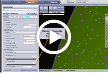
Optimizing Declustring and Edge Detection Settings in Bright Field Imaging
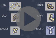
Presenter: Lauren DesRoches, Technical Support Specialist, Nexcelom Bioscience
Assay: Cellometer Software
Instrument: Cellometer Mini,Cellometer Auto 1000,Cellometer Auto 2000
Runtime: 00:48:16
Aired: November 23, 2016
In this user training webinar, Lauren DesRoches, Technical Support Specialist at Nexcelom Bioscience covers how to optimize setting parameters that are useful when working with clumpy samples. Lauren first covers some ways to help optimize your sample preparation to help reduce clumps, and then opens the Cellometer Auto 1000 software to walk users through how to edit the pre-set parameters including edge detection, background contrast and threshold detection on common clumy cell lines including C6, DLD, Ghost and MCF-7.
Optimizing your Exposure Time and Focus Settings
Presenter: Lauren DesRoches, Technical Support Specialist, Nexcelom Bioscience
Assay: Cellometer Software
Instrument: Cellometer K2,Cellometer X1/X2,Cellometer Vision,Cellometer Vision CBA
Runtime: 00:28:33
Aired: October 27, 2016
In this user training webinar, Lauren DesRoches, Technical Support Specialist at Nexcelom Bioscience reviews why, even when using fluorescent staining, achieving a good focus of your sample can be critical to the accuracy of your results. Lauren begins with a review of the Cellometer instruments that use fluorescent imaging and examples of good and bad focused samples. From there, she opens the Cellometer K2 software and walks through how to set and modify the exposure times, while demonstrating how incorrect exposure times can also impact how the Cellometer software analyzes each sample.
How to Perform Apoptosis, Cell Cycle and GFP Tranfsection Assays using the Cellometer Vision CBA
Presenter: Ben Paradis, Technical Support Specialist, Nexcelom Bioscience
Assay: Cell-Based Assays
Instrument: Cellometer Vision CBA
Runtime: 00:44:41
Aired: April 25, 2016
In this user training webinar, Ben Paradis, Technical Support Specialist at Nexcelom Bioscience covers how to perform 3 popular cell-based assays on the Cellometer Vision CBA system. Ben first reviews the software features of the Vision CBA system and also provides training on the FCS Express software, that will generate your histograms and scatter plots for your assays. Ben then covers, one by one, how to perform an Apoptisis assay, Cell Cycle assay, and GFP Transfection Rate assay. Ben concludes by reviewing the data management features available.
How to Measure Concentration and Viability of Primary Cell Samples
Presenter: Dr. Leo Chan, R&D Manager, Nexcelom Bioscience
Assay: Viability
Instrument: Cellometer Auto 2000
Runtime: 00:25:21
Aired: January 23, 2015
In this user training webinar, Dr. Leo Chan, Technology R&D Manager at Nexcelom Bioscience reviews viability assays on the Cellometer Auto 2000. Dr. Chan beings by providing a review of the Auto 2000 system, which includes an overview of the system, software and data management features, to ensure you know what all the features do and how you can use them to your advantage. Dr. Chan explains the assay principle of Acridine Orange/Propidium Iodide before previewing example images of samples that have been counted on the system. Finally, two viability methods are examined: Trypan Blue and Acridine Orange/Propidium Iodide. Dr. Chan examines the benefits of each and helps identify the best method, depending on the cells types being used.
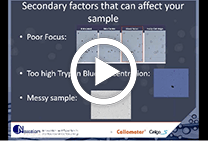
The Importance of Proper Focus When using Bright Field Imaging for Total Cell Count and Viability
Presenter: Dr. Dmitry Kuksin, Assay Development Scientist, Nexcelom Bioscience
Assay: Cell Counting Techniques
Instrument: Cellometer
Runtime: 00:38:07
Aired: October 24, 2014
Have you ever wondered just exactly how the Cellometer software knows what to outline as a cell? Have you questioned whether getting precise focus is really necessary for your sample? In this user training webinar, Dr. Dmitry Kuksin explains the importance of good focus – and why this is important when ising the Bright Field image to determine total cell count and Trypan Blue viability with all our Cellometer systems. He covers how image cytometry works, shows examples of samples that are unfocused, have dim centers, have fuzzy edges and also are properly focused, and explains why an unfocused sample can result in lower viability percentages, or lower total cell counts than you expected.
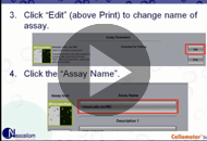
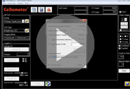
How to Create New and Edit Existing Assays and Cell Types on your Cellometer Vision CBA
Presenter: Stephanie Saldi, Technical Support Specialist, Nexcelom Bioscience
Assay: Cellometer Software
Instrument: Cellometer Vision CBA
Runtime: 00:32:03
Aired: September 26, 2014
In this user training webinar, Stephanie Saldi, Technical Support Specialist at Nexcelom Bioscience will use the Cellometer Vision CBA system and demonstration how users can create new assays and cell types, and also how to edit existing assays and cell types. Stephanie explains the different imaging modes available in the software, and also describes the paramenters for cell type settings. After walking through each step, Stephanie demonstrates how these settings can be changed in a live demonstration.
Trypan Blue Viability on your Cellometer Auto 1000
Presenter: Stephanie Saldi, Technical Support Specialist, Nexcelom Bioscience
Assay: Viability
Instrument: Cellometer Auto 1000
Runtime: 00:16:28
Aired: August 22, 2014
In this user training webinar, Stephanie Saldi, Technical Support Specialist at Nexcelom Bioscience uses the Cellometer Auto 1000 system and demonstrates how to perform the Trypan Blue viability assay. Stephanie reviews the Auto 1000, and the features on the home screen before discussing the protocol for performing Trypan Blue viability assays. She conducts a live demonstration on the Auto 1000, and wraps up the session by demonstrating the data management features.
Yeast Viability Assay on the Cellometer X2
Presenter: Ben Paradis, Technical Support Specialist, Nexcelom Bioscience
Assay: Viability
Instrument: Cellometer X2
Runtime: 00:23:37
Aired: July 25, 2014
Ben begins by reviewing the Cellometer X2 instrument and software features – both the main screen and results screen . He then covers the assay principle for Acridine Orange/Propidium Iodide viability as well as the assay protocol. Ben then opens up the Cellometer X2 software and runs a fresh yeast sample on the system. This webinar concludes with a quick overview of the other assays that are supported by the Cellometer X1/X2 and Cellometer X4 systems including: Yeast Vitality, GFP Transfection, GFP/PI Viability and Cell Cycle analysis.
Primary Cell Viability Analysis using AO/PI and Trypan Blue
Presenter: Ben Paradis, Technical Support Specialist, Nexcelom Bioscience
Assay: Viability
Instrument: Cellometer
Runtime: 00:26:31
Aired: June 24, 2014
In this user training webinar, Ben Paradis, Technical Support Specialist at Nexcelom Bioscience reviews primary cell viability assays. Ben first reviews the assay protocol for Trypan Blue viability staining before exploring a dual fluorescent viability method and looking at why it might be needed. He reviews the assay principle of Acridine Orange/Propidium Iodide before detailing the assay protocol for AO/PI. Finally, two viability methods are examined: Trypan Blue and Acridine Orange/Propidium Iodide. Ben examines the benefits of each method and helps identify which would be the best method, depending on the cells types being used.
How to Create AO/PI Assays on the Cellometer Auto 2000, K2 and Vision
Presenter: Ben Paradis, Technical Support Specialist, Nexcelom Bioscience
Assay: Viability
Instrument: Cellometer Auto 2000,Cellometer K2,Cellometer Vision
Runtime: 00:22:14
Aired: May 23, 2014
In this user training webinar, Ben Paradis, Technical Support Specialist at Nexcelom Bioscience reviews how to perform a viability assay using Nexcelom reagent, Nexcelom ViaStain™ AOPI Staining Solution (Cat# CS2-0106) on the Cellometer Auto 2000, Cellometer K2 and Cellometer Vision instruments. Ben starts by reviewing the assay protocol. Then, he goes through, step-by step, first with the Auto 2000, then with the K2, on creating the assay in the Cellometer software. He also does a live demonstration on the Auto 2000 and Vision. Ben wraps up this session by looking at why you might want to use AOPI for your viability measurements.
How to Perform AO/PI Viability Assays on the Cellometer K2
Presenter: Ben Paradis, Technical Support Specialist, Nexcelom Bioscience
Assay: Viability
Instrument: Cellometer K2
Runtime: 00:28:36
Aired: March 28, 2014
In this user training webinar, Ben Paradis, Technical Support Specialist at Nexcelom Bioscience covers how to perform the AO/PI viability assay on the Cellometer K2, with a special highlight to look at working with hepatocytes. Ben begins with a review of the Cellometer K2 software features before reviewing the AO/PI viability protocol. He reviews in a live demo how to run the assay on the K2, and also covers specific treatment for hepatocytes before wrapping up by reviewing the data management features of the system.
How to Perform Transfection Efficiency and Viability assays on the Cellometer Vision
Presenter: Dr. Dmitry Kuksin, Assay Development Scientist, Nexcelom Bioscience
Assay: Viability
Instrument: Cellometer Vision CBA
Runtime: 00:37:42
Aired: February 28, 2014
In this user training webinar, Dr. Dmitry Kuksin, Assay Development Scientist at Nexcelom Bioscience uses the Cellometer Vision and demonstrations how to perform GFP/RFP transfection efficiency assays, AO/PI viability assays and GFP/PI viability assays. Dr. Kuksin first reviews the software features of the Vision system and software. He then starts the Vision software and runs live samples to demonstration the AO/PI viability assay, GFP/RFP transfection efficiency assay and GFP/PI viability assay. Dr. Kuksin concludes by reviewing the data management features available.
Optimizing Declustering and Edge Detection Settings in Bright Field Imaging
Presenter: Lauren DesRoches, Technical Support Specialist, Nexcelom Bioscience
Assay: Cellometer Software
Instrument: Cellometer
Runtime: 00:30:36
Aired: February 28, 2014
In this user training webinar, Lauren DesRoches, Technical Support Specialist at Nexcelom Bioscience covers how to optimize setting parameters that are useful when working with clumpy samples. Lauren first covers some ways to help optimize your sample preparation to help reduce clumps, and then opens the Cellometer T4 software to walk users through how to edit the pre-set parameters including edge detection, background contrast and threshold detection on common clumy cell lines including C6, DLD, Ghost and MCF-7.