Kinetic measurement of AAV vector transduction efficiencies to optimize assay duration
In this experiment, Celigo was used to image and quantify AAV-GFP transduction efficiencies in 96-well plates for 6 different serotypes at high, mid, and low MOIs over time.
- Target cells are seeded in each well of a 96-well plate and incubated overnight
- Cells are transduced with six types of AAV-GFP vectors at high, medium, and low MOIs and incubated for overnight transduction
- The Celigo plate imager imaged and analyzed in brightfield and green fluorescence to measure the transduction efficiencies at three MOIs for different serotypes of AAV-GFP vectors
- The Celigo analyzed time points at approximately 20, 40, and 60 hours
- The software automatically identifies and segments cells to provide accurate counts of both total cells and GFP+ cells
Determine changes in transduction efficiencies and gene expression levels with time-course measurements of GFP population and fluorescence intensities
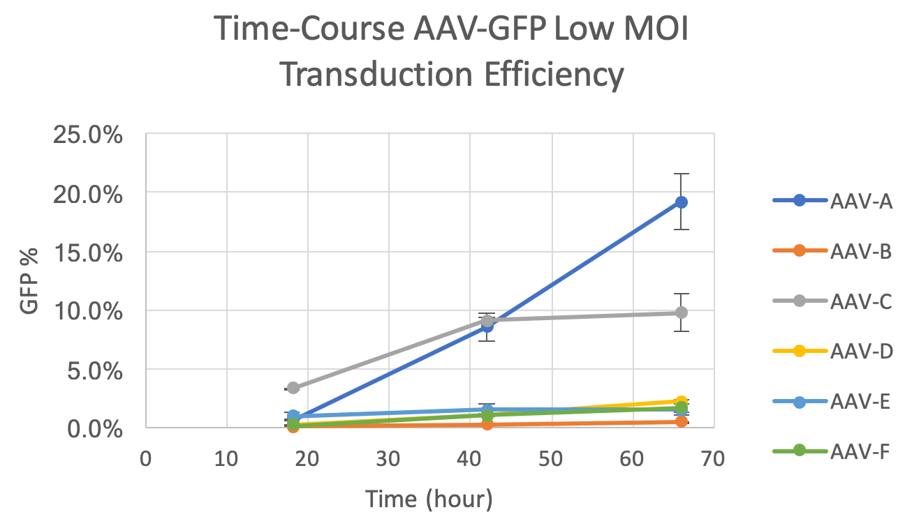
- MOI and time-dependent increases in GFP+ cells were observed for the six different AAV serotypes (Figure 1)
- AAV-C demonstrated the highest efficiency at 20 hours for all three MOIs
- AAV-A and AAV-C had similar percentages of GFP+ percentages at 40 hours
- AAV-A outperformed AAV-C at the final time point
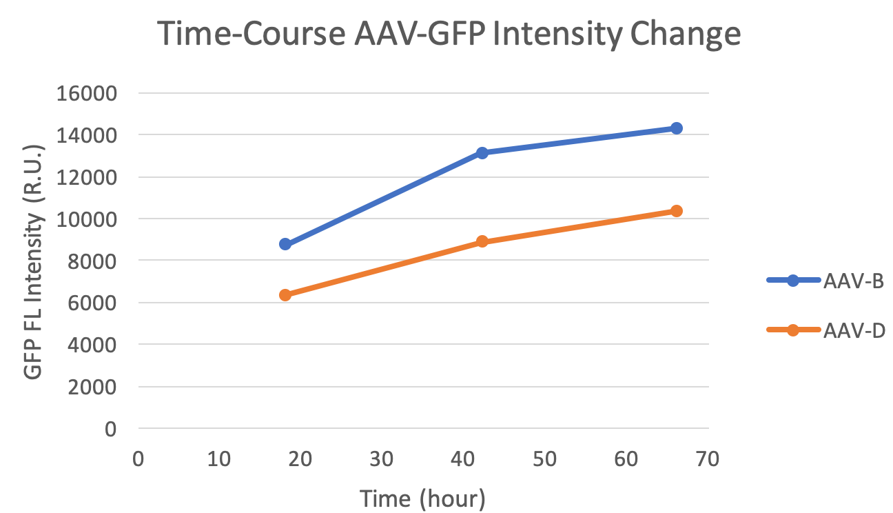
- AAV-B showed higher gene expression over time in comparison to AAV-D (Figure 2)

Figure 1. A time-course plot of six serotypes of AAV vectors at low MOI.

Figure 2. A time-course plot of GFP fluorescent intensities for AAV-B and AAV-D, where AAV-B showed higher gene expression.
Kinetic measurement of AAV vector transduction efficiencies to optimize assay duration
Learn more:
Kinetic measurement of AAV vector transduction efficiencies to optimize assay duration
