Accurate determination of AAV vector transduction efficiencies using an image-based direct cell analyzation method
In mammalian cells, DNA is wound around histones and locked away in the nucleus. The most advantageous keys that fit those locks and facilitate nuclear access are found on viruses. The three types of viral vectors most commonly used in the development of gene therapy products are adeno-associated virus (AAVs, 50%), adenovirus (12%), and lentivirus 11%). The effective viral vector transduction and transgene expression in a specific target cell type is a critical early step in the development process.
Multiple AAV serotypes preferentially infect different cell types. The table below (Table 1) shows which serotypes are best in specific tissues [26]. AAV2 is the most studied. It is also possible to mix and match features of different serotypes. For example, taking a capsid from AAV3 and genome from AAV4 would be denoted as AAV3/4. Developing pseudotyped viruses can change vector tropism or which cell type it infects and can also improve transduction efficiency.
AAV Vector Transduction Efficiencies
| Optimal Serotypes for Specific Tissues | |||||||||
|---|---|---|---|---|---|---|---|---|---|
| Tissue | AAV1 | AAV2 | AAV3 | AAV4 | AAV5 | AAV6 | AAV7 | AAV8 | AAV9 |
| CNS | x | x | x | x | x | x | |||
| Heart | x | x | x | ||||||
| Kidney | x | ||||||||
| Liver | x | x | x | ||||||
| Lung | x | x | x | x | |||||
| Pancreas | x | ||||||||
| Photoreceptor Cells | x | x | x | ||||||
| Retinal Pigment Epithelium | x | x | x | x | x | ||||
| Skeletal Muscle | x | x | x | x | x | ||||
Table 1. Optimal serotypes for specific tissues
Image cytometry enables fluorescence-based, direct cell counting methods to rapidly measure AAV vector transduction efficiencies
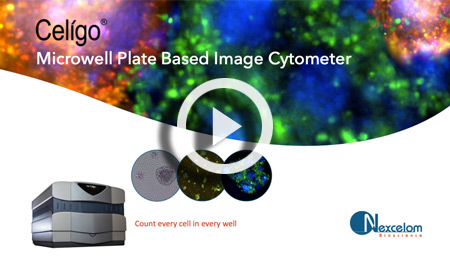
The Celigo Image Cytometer is a plate-based high-throughput system that simultaneously images and directly analyzes cells in standard multi-well plates using brightfield and fluorescence. The Celigo rapidly measures AAV vector transduction efficiencies for the optimization of an AAV vector design.
- Image and analyze 96- and 384-well plates in fluorescence in less than ten minutes per plate
- Direct cell counting method to determine transduction efficiencies using brightfield and fluorescent imaging
- Simultaneously measure transduction efficiencies and fluorescence intensities to quantitatively monitor expression levels
This section provides technical examples of how the Celigo Image Cytometer supports the optimization of AAV vector designs, as well as one example of a lentiviral titration assay.
- Rapidly determine the multiplicity of infection (MOI) dependent AAV vector transduction efficiencies to optimize AAV vector design
- Kinetic measurement of AAV vector transduction efficiencies to optimize assay duration
- Direct measurement of lentiviral dose-dependent transduction efficiencies using GFP fluorescence
The Celigo Image Cytometry system performs high-througput, whole-well imaging and quantitative data in bright field and up to four fluorescent channels for a wide variety of cell-based assays.