Single Cell Per Well Verification
Cell Line Development – Single Cell Detection, Clonal Validation, Transfection
Cell line development process
The process of developing a cell line to produce a specific protein or antibody involves multiple stages, all of which can be greatly aided by Celigo imaging cytometer.
Robotic Integration for Cell Line Development
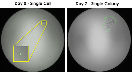
Whole well images: Single cell to single colony

Well with a single cell that grew into a single colony
The Celigo provides an optional robotic API which can be controlled by various automation scheduling software applications. The Celigo is ready for integration with multiple automation partners and can be coupled with robotic arms, automated incubators and liquid handlers.
The Celigo can be used through the whole process of cell line development.
- Compatible with 96-, 384- and 1536-well plates.
- Identify wells with a single colony to avoid the time-consuming and manual identification of clones by eye.
- Measures colony size using bright field and aids the process of selecting wells for clonal expansion.
- Automate cell line generation process with Celigo robotic integration.
Clonal Validation and Expansion
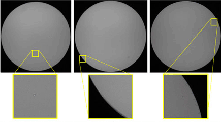
Confirmation of clonality is a critical step in the generation of biopharmaceutical cell lines. With superior bright field image quality, the Celigo cell imaging cytometer reliably visualizes individual cells in a well, even if the cell is adjacent to the well wall. The Celigo allows for verification that a clone originates from a single cell. Typically, limiting dilution is performed in multi-well plates which are then repeatedly imaged on the Celigo over a period of time. When colony growth is seen after a few days of culture, it is then possible to retrospectively confirm that it grew out of a single cell.
Clonal validation on Celigo provides several benefits:
- Fast bright field scanning of multi-well plates (384-well plate can be scanned in <5 min)
- Accurate image segmentation for detection of single cells in a well
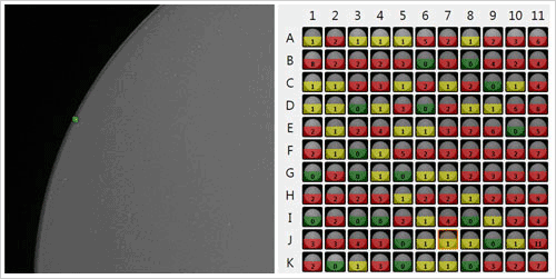
- Heatmap representation of the number of cells/well
- Growth tracking report for monitoring cell growth over time
- Efficient database management of images and data
Clonal Validation on the Celigo Imaging Cytometer

Clonal Expansion on Celigo Imaging Cytometer
Single CHO-S cell to single colony

Zoomed bright field images of a single CHO-S cell expanding into a colony.
Automatic Detection of Single CHO-S Cells on Celigo Imaging Cytometer
Single cell segmentation and heat map

Representative bright field single cell segmentation (left) and resulting heat map of the number of cells per well (right).
