Cellular therapy harnesses the unique properties of cells to restore, repair, or replace damaged tissues or organs in the body. There are various types of cellular therapies, including stem cell therapies, CAR T-cell therapies, and other immunotherapies, that hold great potential in the treatment and prevention of a wide variety of diseases.
The International Organization for Standardization (ISO) has issued guidance on Critical Quality Attributes (CQAs) essential for assessing and approving cellular therapeutic products. These attributes are critical for ensuring the safety, efficacy, and quality of therapeutic products. One CQA that is integral to cellular therapy development is cell viability. Accurate viability measurements can help maintain the quality of the cell sample, which is vital for the feasibility of subsequent downstream experiments.
Choosing reliable viability detection methods
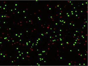
Selecting an appropriate method for measuring cell viability is crucial for consistently obtaining accurate results from a sample. A frequently employed viability detection method involves the use of fluorescent membrane integrity viability stains. These dyes can enter membrane-compromised dead cells, enabling the staining of either cytoplasmic proteins or DNA to produce colorimetric or fluorescent signals. Subsequently, viable and non-viable cells can be distinguished and quantified.
Two commonly-used pairs of fluorescent dyes for assessing live/dead count and total/dead count are acridine orange/propidium iodide (AO/PI) and AO/4′,6-diamidino-2-phenylindole (DAPI) AO/DAPI, respectively. While previous studies have suggested comparable viability results for these staining methods, there have been cases where the methods have indicated noticeable differences. To investigate these disparities further, Huang and colleagues conducted a series of characterization experiments to evaluate the comparability of the two cell viability measurement approaches and identify potential causes for any differences.1
Evaluating cell viability assay methods and measurements
In each experiment, cell viability was determined by pipetting fluorescently stained Jurkat cells into loading wells on Revvity cell counting plates. They were then inserted into a Cellaca® MX high-throughput cell counter for image acquisition and analysis. Following image acquisition, the software calculated the cell count and viability results for cells treated with AO/PI and AO/DAPI.
When the team compared cell viability between the two staining methods, they observed differences in measured cell viability between AO/PI and AO/DAPI (higher). Notably, at higher viability (day 0, viability > 90%) values were more comparable, whereas at lower viability (day 1 to 4, viability < 50%) greater variability was observed. When they investigated whether this difference could be attributed to staining time, they found that AO/PI staining presented consistent results regardless of staining time, whereas AO/DAPI required longer incubation times for accurate measurements.
To better understand the staining dynamics for the two staining methods, the group characterized the cell viability of cell samples across various levels of viable and dead cells. At high viability (100% and 75% cell mixtures) measurements were comparable between the two staining methods. The study shows that as viability decreased (50%, 25%, and 0% cell mixtures), significant differences emerged. Consistent with their earlier results, AO/PI staining maintained consistent viabilities across all mixtures, whereas the AO/DAPI methodology exhibits a continuous decrease in cell viability for all mixtures, except the 100% viable cell samples. Notably, the AO/DAPI viabilities aligned more closely with AO/PI as staining time increased.
The team also conducted a long-term cell proliferation assay to validate viability measurements. This assay reaffirmed that AO/PI consistently identified dead cells, whereas AO/DAPI tended to overestimate viability.
Finally, the researchers compared the two cell viability detection methods using lentivirus-transduced Jurkat and THP-1 cells subjected to antibiotic selection. The ability to accurately measure cell viability throughout this process is critical for preparing cells for downstream screening assays. Overall, the AO/PI staining method showed much higher dead cell concentrations and lower viability than the AO/DAPI, consistent with the results from previous experiments.
Consistent, accurate results at every stage
In summary, the study highlighted the differences between AO/PI and AO/DAPI viability measurement methods, with AO/PI demonstrating more consistent and accurate results across various experimental conditions, independent of staining time and various viability mixtures.
The strategies used in the study can be utilized to compare other techniques and choose an appropriate detection method that enhances the accuracy of evaluating the cell viability of various stages of cellular therapeutic products.
Revvity offers a leading portfolio of automated cell counters, image cytometry systems, and cell counting reagents and consumables, including the Cellaca MX cell counter described in this study. The Cellaca MX utilizes innovative cellular analysis technology for accurate cell sample concentration, viability, and morphology measurements.
References
- Huang Y, Watkins R, Patel S, Pierce M, Franco Nitta C, Qazi H, et al. Practical characterization strategies for comparison, qualification, and selection of cell viability detection methods for cellular therapeutic product development and manufacturing. Journal of Fluorescence. 2023; doi:10.1007/s10895-023-03382-1


Leave A Comment