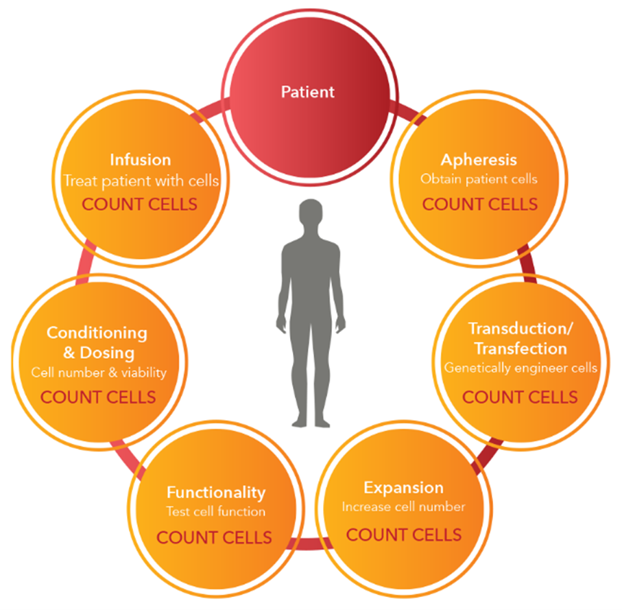
Canine adipose-derived mesenchymal stem cells (cASCs)
When the monolayer of adherent cells reached approximately 70–80% confluence; trypsinization for cell splitting was performed using trypsin-EDTA solution (0.05%/0.02%, Biochrom AG, Berlin, Germany). After centrifugation, the cell pellets were subcultured at a concentration of 3 × 10⁵cells/mL medium on 25cm2 tissue culture flasks containing DMEM/10% FCS. Cell counting was carried out by an automatic cell counter (Cellometer Nexcelom Bioscience, Lawrence, USA).
The viable cell percentage from the harvested cells was calculated after trypan blue staining by an automatic cell counter (Cellometer Nexcelom Bioscience, Lawrence, USA). The cell concentration used for the proliferation assay was adjusted to 7×10³ cells/100μL medium.

Leave A Comment