It’s White Paper Wednesday! Read our featured white paper: Concentration and Viability Measurement of Canine Stromal Vascular Fractions using Cellometer Vision
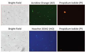 Cell concentration and viability of SVF preparations are usually determined by standard hemocytometer methods that are prone to considerable error since the operator must make judgments between actual cells versus “debris”. To address that problem, we employed Cellometer image cytometry to perform both bright-field and fluorescence-based cell concentration and viability measurements [12]. Here, we validated this method for SVF analysis. First, the imaging parameters were optimized by measuring five adipose SVF samples. Next, the concentration and viability of three freshly prepared SVF cell samples were measured and compared using hemocytometer, flow cytometer, and image cytometer methods using trypan blue (TB) and a mixture of Hoechst 33342 and propidium iodide (HO/PI). In addition, a mixture of acridine orange and propidium iodide (AO/PI) was used to measure concentration and viability for the image cytometry method for comparison to HO/PI. The results show comparable concentration measurements amongst the detection methods used, and show that automated image-based cytometry can be used to efficiently generate accurate SVF measurements.


Leave A Comment