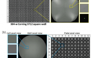
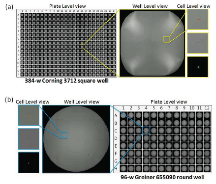
Using the Cellometer Vision Image Cytometer for Immunophenotyping
It's White Paper Wednesday! Read our featured white paper: Using the Cellometer Vision Image Cytometer for Immunophenotyping By developing fluorescent-based assays to immunophenotype cells, the Cellometer imaging cytometry can provide a quick, simple, and inexpensive alternative for biomedical research, which may be beneficial for smaller research laboratories and clinics. In this publication, we demonstrate an immunophenotyping assay to detect percentages of lymphocyte populations in the spleen and thumus via cell surface markers CD4, CD8, B220, and CD5 with Cellometer imaging cytometry as an alternative to flow cytometry. The data obtained by Cellometer were compared to those from conventional flow cytometry [...]