Transcript: Assessment of Mitochondrial Membrane Potential using Image-Based Cytometry
Cellometer Vision CBA is a simple, image-based instrument optimized for the analysis of fluorescent cell-based assays.
Mitochondrial membrane potential is often used to screen for early-stage cell death. To run this experiment, Jurkat cells were incubated with a JC-1 staining solution for 30 minutes at 37 degrees C. Following incubation, 20µl of sample was pipetted into the Cellometer counting chamber. The chamber was inserted into the Vision CBA instrument.
The Mitochondrial Membrane Potential (JC-1) Assay is selected from the drop-down menu. The specific Sample ID is entered, then click count.
In less than 3 minutes, the Vision CBA takes multiple cell images and analyzes the images based on pre-set parameters for the Mitochondrial Membrane Potential Assay selected.
When cell imaging and counting is complete, the initial results table displays total cells counted, concentration, and mean diameter.
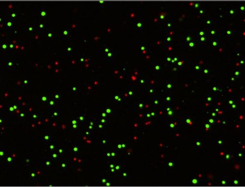
The brightfield image can be viewed to verify cell morphology and identify cell clumps. The JC-1 dye accumulates in the mitochondria of healthy cells as aggregates which are fluorescent red in color. Upon onset of cell death, the mitochondrial membrane potential is compromised and the JC-1 dye remains in the cytoplasm in a monomeric form that fluoresces green.
The export function is used to generate results in FCS Express 4 Software using optimized Vision CBA data layouts. Mitochondrial membrane potential is displayed as a colorized scatter plot showing % compromised and % healthy cells based on red and green fluorescence. The gating can be manually optimized. Associated data tables update automatically to reflect revised percentages.
Users can choose to print a second report containing both brightfield and fluorescent images. Data tables and cell images can be easily exported for use in presentations and publications.
After removing the disposable Cellometer cell-counting chamber, the Vision CBA is ready to analyze the next sample. No washing or instrument set-up is required.
Cellometer Vision CBA offers researchers a simple, image-based option for cell-based assays. Visit www.nexcelom.com or contact Nexcelom today to schedule an on-site demonstration with an experienced applications specialist.


Leave A Comment