| Purpose | To provide an automated solution that can provide images of multicellular tumor spheroids (MCTS) and report MCTS diameter in a 96-well format |
| Current Method(s) | Microscopy |
| Target Cell Type | NCI-H460, MiaPaca |
| Experiment Plan | Scan two plates daily to measure the size of multicellular tumor spheroids under different drug concentrations in hypoxic or normoxic conditions |
| Hypothesis | Using the bright field imaging, the Celigo will rapidly provide multicellular tumor spheroid images and diameters of treated MCTS in a 96-well plate |
Celigo Setup
| Plate Type | Corning™ CLS70007 (ULA plate) – 96 well plate |
| Scan Channels | Bright field |
| Resolution | 3 micron/pixel |
| Scan Area | Whole well |
| Analysis Method | Tumorsphere 1 |
| Scan Frequency | Daily |
| Scan Time | 3 minutes |
Assay Protocol and Plate Setup
Goal:
Image and measure the diameter of multicellular tumor spheroids (MTSs) at different drug concentrations that were incubated at either normoxic or hypoxic conditions. Ultimately determine, if incubation conditions (normoxic, and hypoxic) play a role in the reduction of MTS size when treated at different drug concentrations.
Protocol:
Cell Preparation
- Seeded 2,500 NCI-H460 cells/well in a ULA 96-well plates (see plate map below)
- Seeded 1,250 MiaPaca cells/well in a ULA 96-well plates (see plate map below)
- On day 4, treated formed MTSs with drug X or vehicle control
- Drug X in [ µM ] was serially diluted 1/3 from 10 µM to 0.0045 µM concentration
- Placed one plate in hypoxic conditions and one plate at standard growth conditions
Plate map:

Data Collection
- On day 4, after adding the drug at different concentrations, the plates were imaged and data collected for both the hypoxia and normoxia plates
- The plates were again imaged 4 days later. Post-treatment, one plate was at hypoxic conditions for 4 days and the second plate was normoxic conditions
- The plates were scanned in Celigo using Tumorsphere 1 (bright field) assay
Data Analysis
- The images were analyzed by using Tumorsphere 1 application to identify the MCTS in the well
- The well mask was reduced to the 95th percentile to eliminate edge effects
- Several wells had multiple multicellular tumor spheroids. In those instances, each spheroid was identified by the Celigo software
Results
- NCI-H460 multicellular tumor spheroids showed a noticeable decrease in spheroid size at high drug concentration
- NCI-H460 multicellular tumor spheroids showed a decrease in spheroid size from 780 µm at 3 µM drug concentration to 750 µm at normoxic and 720 µm at hypoxic conditions at 10 µM.
- MiaPaca multicellular tumor spheroids showed no change in spheroid diameter between hypoxic and normoxic conditions for control or drug-treated samples.
- Image and graphed data below represents NCI-H460 and MiaPaca multicellular tumor spheroids treated with Drug X in a dose-dependent manner and incubated at normoxic and hypoxic environment for four days before imaging on the Celgio.
Day 4 post-treatment with Drug X at Normoxia NCI-H640 at 2,500 and MiaPaca at 1,250 cells/well
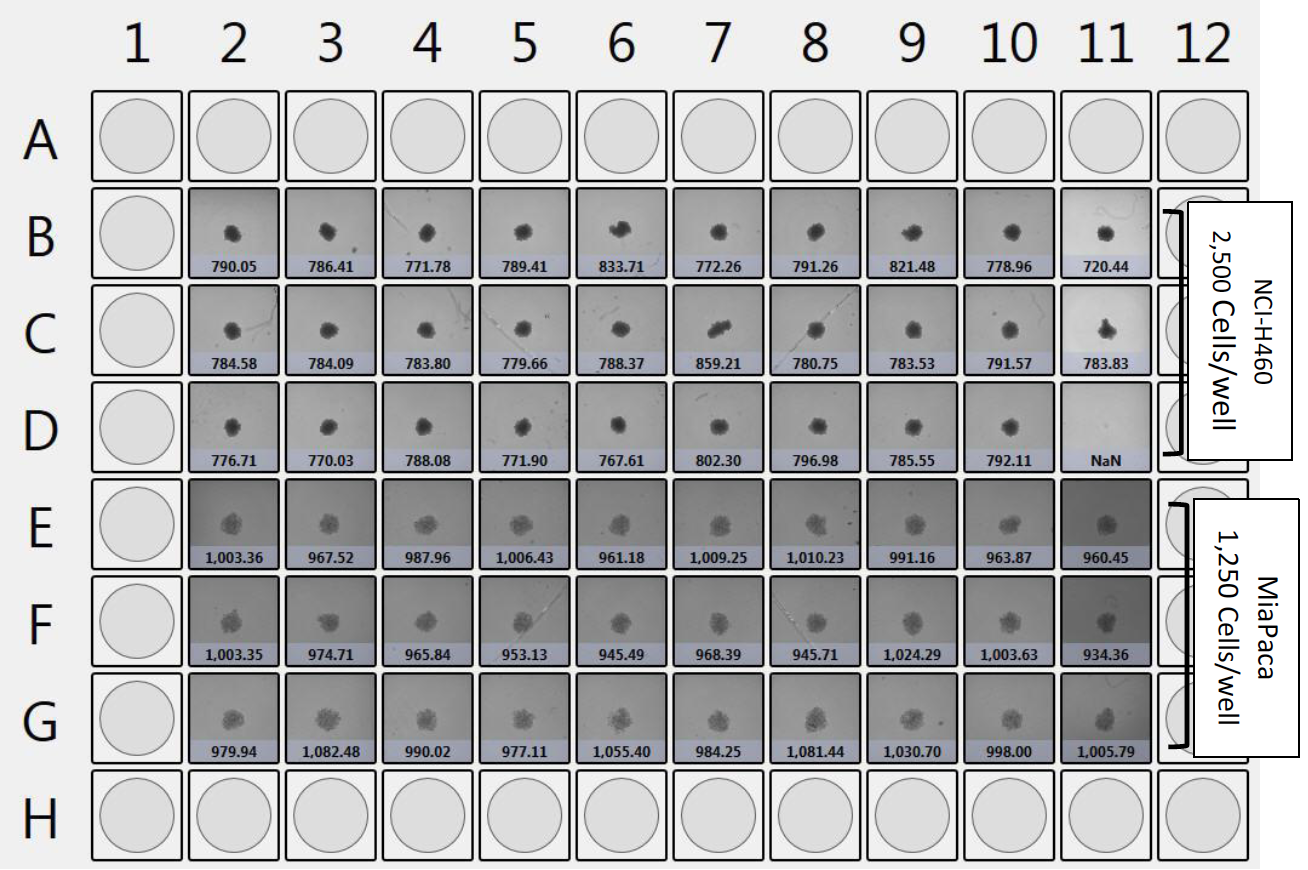
- The entire plate was scanned in 3 minutes and bright field thumbnails are shown for each well.
- At the bottom of the well picture is the Celigo measured diameter in (microns) for each identified spheroid.
Day 4 post-treatment with Drug X at Hypoxia NCI-H640 at 2,500 and MiaPaca at 1,250 cells/well
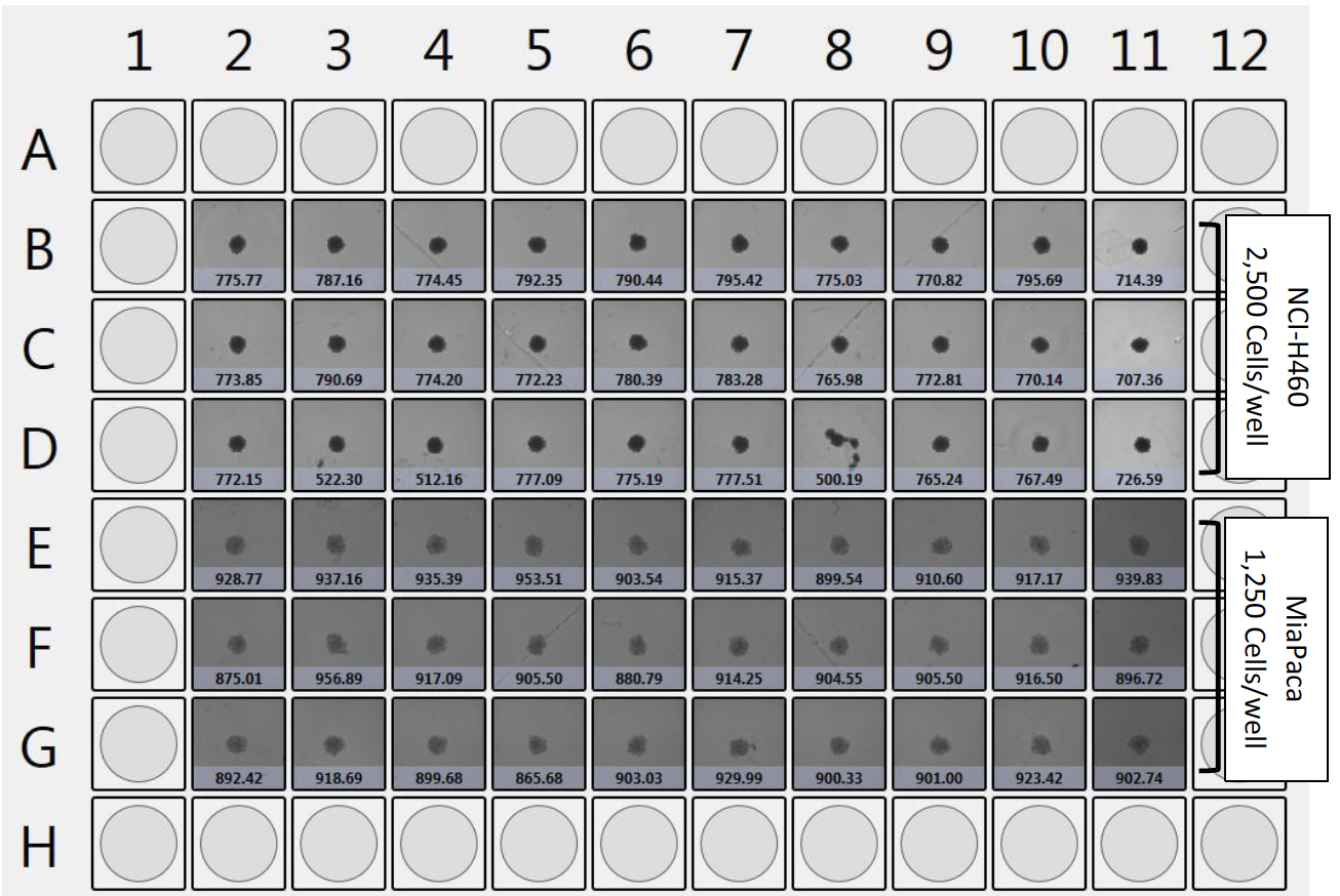
- The entire plate was scanned in 3 minutes and bright field thumbnails are shown for each well.
- At the bottom of the well picture is the Celigo measured diameter in (microns) for each identified spheroid.
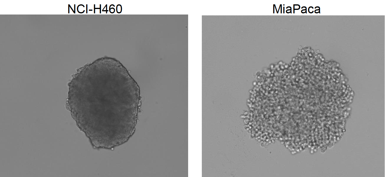
Celigo captured representative bright field images of non-drug treated NCI-H460 and MiaPaca multicellular tumor spheroids at normoxic condition on day 0
NCI-H460 and MiaPaca multicellular tumor spheroid measurements four days after drug treatment and incubation at normoxic and hypoxic conditions
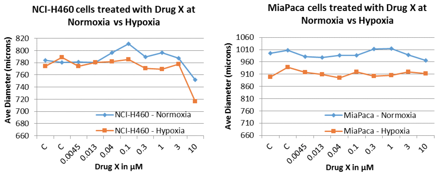
- NCI-H460 and MiaPaca were plated at 2,500 and 1,250 cells per well treated with a serially diluted Drug X and incubated for 4 days at either hypoxic or normoxic conditions
- NCI-H460 multicellular tumor spheroids showed a noticeable decrease in spheroid size at 10 µM drug concentration at both normoxic and hypoxic conditions.
- The sizes of MiaPaca multicellular tumor spheroids remained the same throughout the experiment at both normoxic and hypoxic conditions as well as at different drug concentrations.
Conclusion
- Using the 96-well U-bottom ULA plates, we successfully captured images of MCTS and analyzed the data using the Celigo instrument.
- The entire 96-well plate was imaged in 3 minutes. The short scan time significantly increases the throughput during an experiment that has multiple plates.
- After 4 days of drug treatment and incubation at either normoxic or hypoxic conditions, spheroid diameters were measured and automatically reported by the Celigo software. No additional software is required for image processing of multicellular tumor spheroid diameters