Perform image-based GFP analysis WITHOUT all of the set-up, maintenance, and data manipulation required for flow.
Measure GFP Expression Efficiency Using Image Cytometry —
In just a few clicks, Cellometer Vision CBA generates brightfield and fluorescent cell images, and detailed data reports.
- Detection of GFP expression to quantify the transfection/transduction efficiency.
- Single assay for measuring GFP expression and viability
- Measurement of other fluorescent proteins
Detection of GFP Expression to Quantify the Transfection/Transduction Efficiency
Quantitative measurement of GFP expression using Cellometer
- Load only 20 µl sample into counting chamber
- Capture and analyze brightfield and fluorescent cell images automatically
- Data read-out includes cell counts, concentrations, and percentage of GFP positive cells
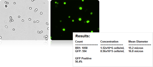
Cellometer cell images and results
Single Assay for Measuring GFP Expression and Viability in Mouse Embryonic Stem Cells
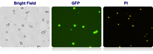
Bright Field, GFP and PI cell images from Cellometer
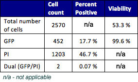
Cellometer data
- Stain Sample with propiduim iodide
- Monitor the viability of the culture during GFP expression
- Determine the viability of GFP expressing cells
Measurement of Other Fluorescent Proteins
Cellometer can measure other fluorescent proteins including dared, YFP, CFP, tdTomato and Saphire.
Customer Publications Using Cellometer for GFP Detection »
Visit the Vision CBA Image Cytometry System Web Page »
Advantages of Image Cytometry:
- No washing. No clogging. No daily calibration
- Bright field and fluorescent cell images for verification of counted cells
- Simple procedure with pre-defined instrument settings and data output
- Small, 20µl sample size
- Non-fluidic instrument … maintenance-free
View the Complete Listing of Cellometer Cell-Based Assay Kits »


Leave A Comment