A Rapid Alternative Method for Cell Cycle Analysis Using Cellometer Vision
Leo L. Chan, Xuemei Zhong, Jean Qiu, Peter Y. Li, and Bo Lin
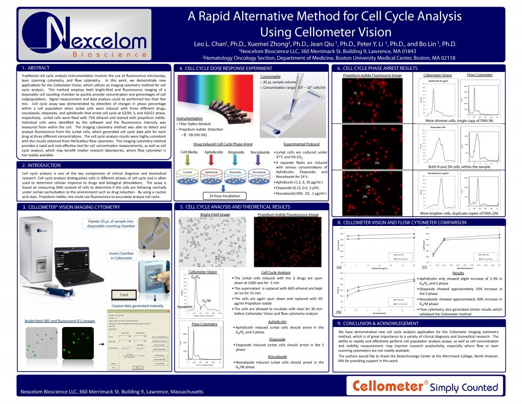
Traditional cell cycle analysis instrumentation involves the use of fluorescence microscopy, laser scanning cytometry, and flow cytometry. In this work, we demonstrate new applications for the Cellometer Vision, which utilizes an imaging cytometry method for cell cycle analysis. This method employs both bright-field and fluorescence imaging of a disposable cell counting chamber to quickly provide concentration and percentages of cell subpopulations. Signal measurement and data analysis could be performed less than five
min. Cell cycle assay was demonstrated by detection of changes in phase percentage within a cell population when Jurkat cells were induced with three different drugs, nocodazole, etoposide, and aphidicolin that arrest cell cycle at G2/M, S, and G0/G1 phase, respectively. Jurkat cells were fixed with 75% ethanol and stained with propidium iodide. Individual cells were identified by the software and the fluorescence intensity was measured from within the cell. The imaging cytometry method was able to detect and analyze fluorescence from the Jurkat cells, which generated cell cycle data plot for each drug at three different concentrations. The cell cycle analysis results were highly consistent with the results obtained from FACScalibur flow cytometer. This imaging cytometry method provides a rapid and cost-effective tool for cell concentration measurements, as well as cell cycle analysis, which may benefit smaller research laboratories, where flow cytometer is not readily available.