Automated Morphometric Analysis of Mammospheres: Characterization of Breast Cancer Drugs
Olivier Dery, Sarah Kessel, Kristi Hohenstein Elliott, and Fredrik Kamme
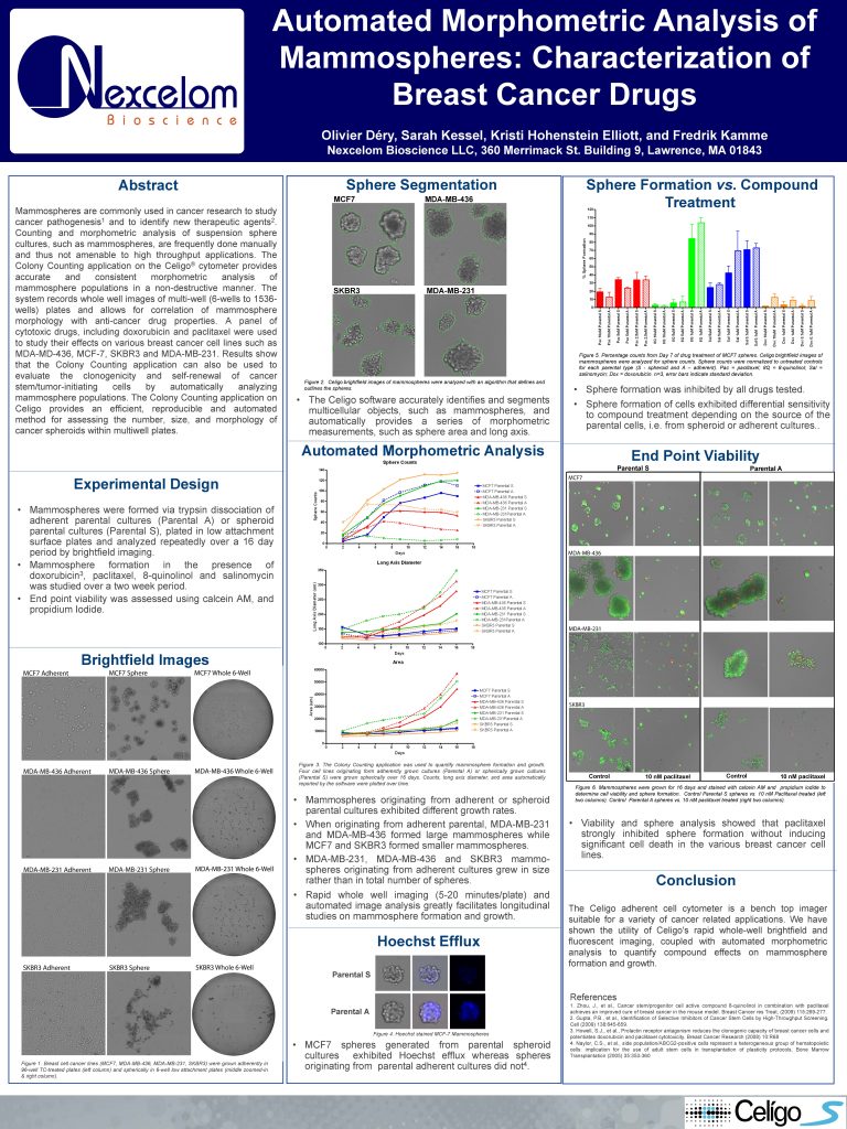
Mammospheres are commonly used in cancer research to study
cancer pathogenesis1 and to identify new therapeutic agents2.
Counting and morphometric analysis of suspension sphere
cultures, such as mammospheres, are frequently done manually
and thus not amenable to high throughput applications. The
Colony Counting application on the Celigo® cytometer provides accurate and consistent morphometric analysis of mammosphere populations in a non-destructive manner. The system records whole well images of multi-well (6-wells to 1536-wells) plates and allows for correlation of mammosphere
morphology with anti-cancer drug properties. A panel of cytotoxic drugs, including doxorubicin and paclitaxel were used to study their effects on various breast cancer cell lines such as MDA-MD-436, MCF-7, SKBR3 and MDA-MB-231. Results show that the Colony Counting application can also be used to evaluate the clonogenicity and self-renewal of cancer stem/tumor-initiating cells by automatically analyzing mammosphere populations. The Colony Counting application on Celigo provides an efficient, reproducible and automated method for assessing the number, size, and morphology of
cancer spheroids within multiwell plates.

