Image Cytometry-Based High-Throughput Cell Concentration and Viability Detection Method using AOPI Fluorescent Stains
Leo L. Chan, Tim Smith, Kendra A. Kumph, Dmitry Kuksin, Sarah Kessel, Olivier Déry, Scott Cribbes, Ning Lai and Jean Qiu
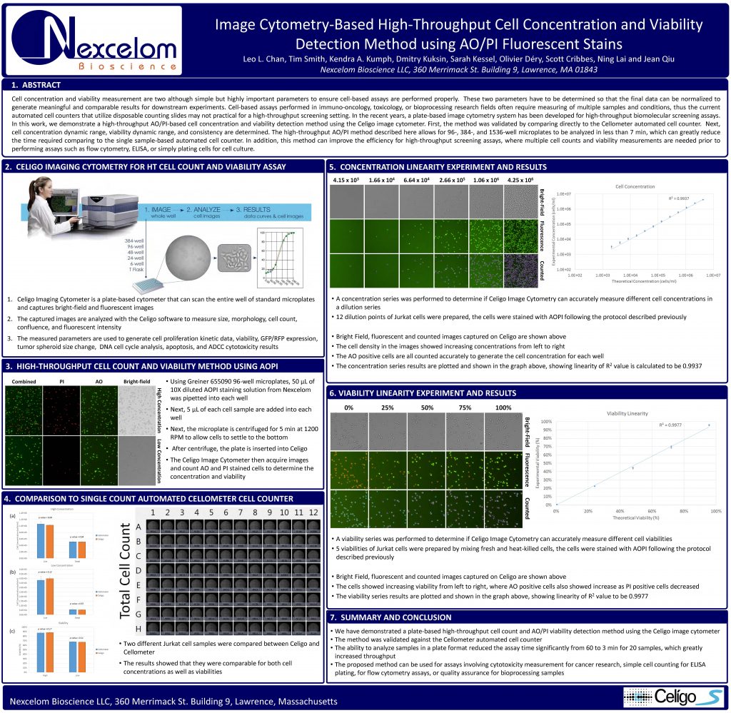
Cell concentration and viability measurement are two although simple but highly important parameters to ensure cell-based assays are performed properly. These two parameters have to be determined so that the final data can be normalized to generate meaningful and comparable results for downstream experiments. Cell-based assays performed in immuno-oncology, toxicology, or bioprocessing research fields often require measuring of multiple samples and conditions, thus the current automated cell counters that utilize disposable counting slides may not practical for a high-throughput screening setting. In recent years, a plate-based image cytometry system has been developed for high-throughput biomolecular screening assays.
In this work, we demonstrate a high-throughput AO/PI-based cell concentration and viability detection method using the Celigo image cytometer. First, the method was validated by comparing directly to the Cellometer automated cell counter. Next, cell concentration dynamic range, viability dynamic range, and consistency are determined. The high-throughput AO/PI method described here allows for 96-, 384-, and 1536-well microplates to be analyzed in less than 7 min, which can greatly reduce
the time required comparing to the single sample-based automated cell counter. In addition, this method can improve the efficiency for high-throughput screening assays, where multiple cell counts and viability measurements are needed prior to performing assays such as flow cytometry, ELISA, or simply plating cells for cell culture.

