Morphological observations using image cytometry for the comparison of trypan blue and fluorescence-based cellular viability
Leo L. Chan, Dmitry Kuksin, Daniel J. Laverty, Jean Qiu, and Bo Lin
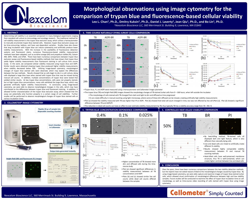
Determining cell viability is an essential component in many biological experiments ranging from standard cell culture to immunology and oncology research. The traditional method for cell viability measurement is the trypan blue exclusion assay, which utilizes a hemacytometer to manually enumerate trypan blue stained cells. However, trypan blue exclusion assays can
be time-consuming, tedious, and have user-dependent variation. Studies have also shown that long incubation with trypan blue can induce cytotoxicity and artificially produce lower viability measurements. With the increase in the availability of fluorescence detection systems and fluorescent stains, numerous fluorescence-based viability measurement methods have been adopted, such as acridine orange (AO) and propidium iodide (PI), Calcein AM, DAPI, 7AAD, and CFDA. There have been numerous comparisons between trypan blue exclusion assays and fluorescence-based viability methods that have shown that trypan blue yields higher viability measurements than fluorescent staining in cell culture time course study. In this work, we compared cellular viability measured using trypan blue and AOPI. Similar results were obtained showing trypan blue produced higher viability measurements when viability decreased below 70%. Utilizing image-based cytometry, morphological changes of trypan blue stained cells were observed, which may explain the differences between the two methods. Results showed that as cells begin to die in a cell culture, dying cells displayed a large blue haze when stained with trypan blue that may be missed during manual cell counts. In addition, multiple concentrations of trypan blue were tested that yielded similar results. At low trypan blue concentrations, cells were not properly stained, but at high trypan blue concentrations, many cells exhibited the large blue haze, which may
generate artificially higher viability measurements. In conclusion, using image-based cytometry, we were able to observe morphological changes in the cells, which may have contributed to the differences between trypan blue and fluorescent staining. In addition, it allowed further characterization of the trypan blue exclusion assay, which showed that the assay can potentially only function properly in a certain range of cell viabilities. Further studies will be performed to observe morphological changes of trypan blue stained cells at various incubation times

