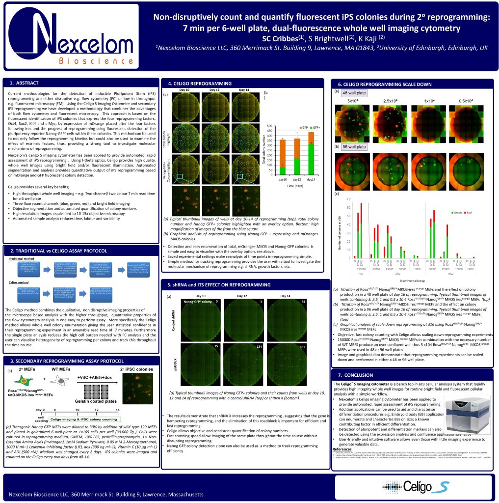
Non-disruptively count and quantify fluorescent iPS colonies during 2° reprogramming: 7 min per 6-well plate, dual-fluorescence whole well imaging cytometry
Scott Cribbes, Sarah Brightwell, and K Kaji
Current methodologies for the detection of inducible Pluripotent Stem (iPS)
reprogramming are either disruptive e.g. flow cytometry (FC) or low in throughput e.g. fluorescent microscopy (FM). Using the Celigo S Imaging Cytometer and secondary iPS reprogramming we have developed a methodology that combines the advantages of both flow cytometry and fluorescent microscopy. This approach is based on the
fluorescent identification of iPS colonies that express the four reprogramming factors, Oct4, Sox2, Klf4 and c-Myc, by expression of mOrange placed after the four factors following ires and the progress of reprogramming using fluorescent detection of then pluripotency reporter Nanog-GFP+ cells within these colonies. This method can be used to not only follow the reprogramming kinetics but could also be used to examine the
effect of extrinsic factors, thus, providing a strong tool to investigate molecular mechanisms of reprogramming. Nexcelom’s Celigo S imaging cytometer has been applied to provide automated, rapid
assessment of iPS reprogramming . Using f-theta optics, Celigo provides high quality, whole well images using bright field and/or fluorescent illumination. Automated segmentation and analysis provides quantitative output of iPS reprogramming based on mOrange and GFP fluorescent colony detection.
Celigo provides several key benefits;
• High throughput whole well imaging – e.g. Two channel/ two colour 7 min read time
for a 6 well plate
• Three fluorescent channels (blue, green, red) and bright field imaging
• Objective segmentation and automated quantification of colony numbers
• High resolution images equivalent to 10-15x objective microscopy
• Automated sample analysis reduces time, labour and variability