Surface Marker Based Direct Cell Concentration Measurements Using a High Sensitivity Imaging Cytometry Method
Leo L. Chan, Bo Lin, Peter Y. Li, and Jean Qiu
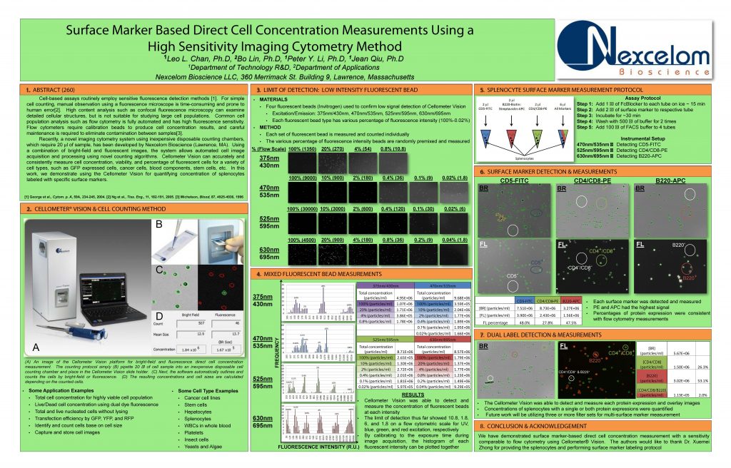
Cell-based assays routinely employ sensitive fluorescence detection methods [1]. For simple cell counting, manual observation using a fluorescence microscope is time-consuming and prone to human error
[2]. High content analysis such as confocal fluorescence microscopy can examine detailed cellular structures, but is not suitable for studying large cell populations. Common cell population analysis such as flow cytometry is fully automated and has high fluorescence sensitivity. Flow cytometers require calibration beads to produce cell concentration results, and careful
maintenance is required to eliminate contamination between samples
[3]. Recently, a novel imaging cytometry system using inexpensive disposable counting chambers, which require 20 μl of sample, has been developed by Nexcelom Bioscience (Lawrence, MA). Using a combination of bright-field and fluorescent images, the system allows automated cell image
acquisition and processing using novel counting algorithms. Cellometer Vision can accurately and consistently measure cell concentration, viability, and percentage of fluorescent cells for a variety of cell types, such as GFP expressed cells, cancer cells, blood components, stem cells, etc. In this
work, we demonstrate using the Cellometer Vision for quantifying concentration of splenocytes labeled with specific surface markers