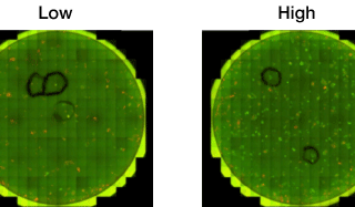
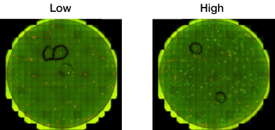
Non-Disruptive Quantification of 2° Reprogrammed iPS Colonies Using Celigo Imaging Cytometer
Celigo imaging cytometer has been applied to provide automated, rapid assessment of iPSC reprogramming.
Celigo imaging cytometer has been applied to provide automated, rapid assessment of iPSC reprogramming.
It's White Paper Wednesday! Read our featured white paper: Automated Method for Determination of Infectious Dose (TCID50) using Celigo Imaging Cytometer Nexcelom's Celigo imaging cytometer has been applied to provide automated, rapid assessment of viral infectivity in a range of plate formats [4]. Using f-theta optics, Celigo provides high quality, whole-well images using bright field and/or fluorescent illumination. Automated segmentation and analysis provide quantitative and objective output of CPE based on characteristic changes to the host cell monolayer. Download our white paper »
It's White Paper Wednesday! Read our featured white paper: Characterization of Breast Cancer Drugs via Mammosphere Morphometric Analysis Using Celigo Imaging Cytometer A panel of cytotoxic drugs, including doxorubicin and paclitaxel were used to study their effects on various breast cancer cell lines such as MDA-MD-436, MCF-7, SKBR3 and MDA-MB-231. Results show that the Colony Counting application can also be used to evaluate the clonogenicity and self-renewal of cancer stem/tumor-initiating cells by automatically analyzing mammosphere populations. The Colony Counting application on Celigo provides an efficient, reproducible and automated method for assessing the number, size, and morphology of cancer spheroids within [...]
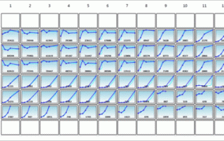
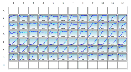
Generate growth curves over time and monitor cell counts and confluence at the individual well level.