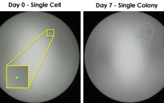
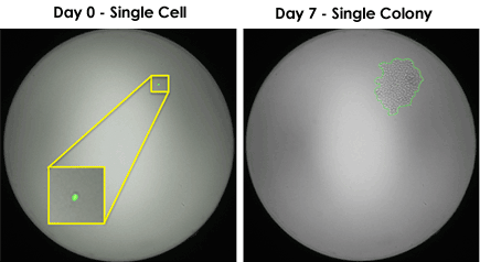
Celigo Provides Image-Based Proof of Single-Cell Clonality
The FDA identifies clonality as one of the most crucial steps in guaranteeing cell line quality and safety. The Celigo Image Cytometer makes single cell sorting easy.
The FDA identifies clonality as one of the most crucial steps in guaranteeing cell line quality and safety. The Celigo Image Cytometer makes single cell sorting easy.
Download our Poster Abstract: Cancer immunotherapy has been gaining momentum in the field of cancer research. Specifically, Chimeric Antigen Receptor (CAR) T cell technology have introduced new methods to combat cancer. Direct cell-mediated cytotoxicity assays are required to assess the killing capability of the engineered CAR T cells. Traditionally, these assays are conducted by measuring the amount of released Chromium, calcein AM, or LDH molecules after the target cancer cells are killed with CAR T cells. These methods require a large amount of target cells which may not be ideal when working with donor primary samples. Additionally, they cannot [...]
Celigo playing an important role in high throughput, high-resolution image acquisition for DNA damage detection. The Celigo S captures 16 images of each well that are then stitched into a single representation Open access article: Next generation high throughput DNA damage detection platform for genotoxic compound screening
By directly imaging and counting every cell in a well over a course of a drug treatment, the Celigo can perform the cytotoxicity assays in a label-free format.
Lawrence, Massachusetts – September, 2016 - Nexcelom Bioscience, a leading provider of cell counting and analysis products for biomedical research and the biopharma industry, announced it has released the addition of a 5th channel available as an option on the Celigo image cytometer. The Celigo is a bench-top image cytometry system providing whole-well imaging and quantitative data, through image analysis for demanding cell-based analytical applications. Previous configurations allowed bright field only imaging, or bright field imaging plus 3 fluorescent channels. This new option of bright field plus 4 fluorescent (red, green, blue and far-red) channels will allow researchers more capabilities [...]
Emily Whitehead, cancer survivor, holding Nexcelom cell counting sheep. Thanks to the lifesaving T cell therapy clinical trial at Children’s Hospital of Philadelphia, Emily Whitehead is now two years cancer free. Diagnosed with acute lymphoblastic leukemia (ALL) right after her 5th birthday, doctors discovered that Emily’s leukemia was particularly resistant to chemotherapy, as are roughly 15% of the total number of ALL cases. After two recurrences of the disease, Emily’s parents enrolled her in a clinical trial for CTL019, an experimental therapeutic using a patient’s own reprogrammed T cells to eliminate the cancer cells. Now 9 years old, [...]
Recent publications have suggested that using 3D tumor spheroids is a more predictive model for preclinical research. Nexcelom Bioscience has developed a standardized microplate method for rapid generation, imaging and analysis of 3D tumor spheroids using the Celigo image cytometer. 40 cancer cell lines' ability to form spheroids, optimal seeding densities and culture conditions have been established. Protocols measuring tumor growth, viability, migration and invasion have been utilized by many researchers for routine preclinical drug studies. If you are interested to learn more about how your research might benefit from working with 3D tumor spheroids, this is a great place to start! [...]
Celigo Imaging Cytometer demonstrated the utility of automation in the development and monitoring of new CHO-based cell lines for increasing efficiency in cell line development.
It's White Paper Wednesday! Read our featured white paper: A Rapid and Label-Free In Situ Assay Method for Cell Proliferation and Drug Toxicity using Celigo Imaging Cytometer In this study, Celigo was used to screen a compound library for effects on cell proliferation in adherent and non-adherent cell lines. Human lung carcinoma (A549) and promyelocytic leukemia (HL-60) cells were treated with a panel of compounds to inhibit proliferation. Finally, the Celigo system used image-based analysis to measure changes in cell morphology upon compound treatment. These data indicate that certain anti-proliferative compounds can have secondary effects on cell health or physiology, [...]
It's White Paper Wednesday! Read our featured white paper: 3D Tumor Spheroid Analysis Method for HTS Drug Discovery using Celigo Imaging Cytometer U87MG cells were used to create tumorspheres in 384-well plates that were subsequently analyzed by imaging. The data illustrate that reproducible 3D spheroids can be formed in 384-well plates. Fluorescent viability studies were carried out with the imager using pixel intensity analysis. Moreover, the assay was validated for drug screen using various drug compounds that have shown anti-proliferative effects. Together, these data demonstrate that the tumorsphere formation assay can be developed, validated and used for high-throughput anti-cancer compound [...]