The hemocytometer has been an essential tool for hematologists, medical practitioners, and biologists for over a century. Depending on where it is being used, the word has multiple spellings such as hemacytometer, hemocytometer, haemacytometer, or haemocytometer, but for consistency purposes, the word “hemocytometer” will be used in this review. The prefix “hema”, “hemo”, “haema”, or “haemo” means blood, while “cytometer” meant a device to measure cells. The device was initially used by medical practitioners to analyze patient blood samples, which was the initial spark that created the field of hematology. The hemocytometer has gone through a series of major development in the 1800s and early 1900s. The modern-day hemocytometer utilizes a double-chamber format and counting grids developed by O. Neubauer. This format allows the user to perform two cell counts for each sample without having to clean it. For a complete historical review, please download our white paper.
The hemocytometer has been used to count cells ranging from algae, yeast, cancer cells, stem cells, blood cells, even parasites and spores. Although a variety of automated cell counting instruments have been developed, the current golden standard that researchers fall back on is still manual counting with hemacytometer.
Manual Cell Counting with Hemocytometer
Step 3. Manually Count Cells in Sample
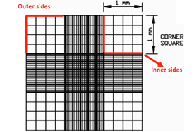
Focus both onto the grid pattern and the cell particles, and count the total number of cells found in 4 large corner squares.
If cells are touching the 4 perimeter sides of a corner square, only count cells on 2 sides, either the 2 outer sides or 2 inner sides.
Step 4. Cell Calculations & Disposal
Multiply the dilution factor by the total number of cells, divide by the # of corner squares counted, and multiply by 104 to obtain cell concentration (cells/ml).
Clean hemocytometer and glass coverslip with 70% EtOH.




Leave A Comment