What is GFP?
Green Fluorescent Protein (GFP) is a 26.9 kDa protein first identified in crystal jellyfish, Aequorea victoria. It was discovered that when exposed to blue or ultraviolet light the protein fluoresces green. After GFP was first expressed in E. coli in 1994 it was soon confirmed that GFP can also be successfully expressed in other organisms as well. Since then, not only have many fluorescent proteins of different colors been generated, but their function is enhanced to provide a faster and stronger fluorescent signal.
GFP Applications
- GFP is often used as a reporter of gene or protein expression. By detecting GFP expression it is possible to quantify the transfection/transduction efficiency.
- By staining the cells with propidium iodide we can monitor the viability of the culture during GFP expression.
- In cultures that are co-transduced with GFP and RFP, the Cellometer has the capability to capture, analyze, and report the population of GFP positive, RFP positive, or dual positive.
Acquiring GFP Expression Efficiency Using Cellometer
With the Cellometer Vision CBA, just 20µl of sample is added to the Cellometer Counting Chamber. Imaging and analysis of GFP expression is completed in less than 60 seconds. Bright field and fluorescent cell images can be viewed to check cell morphology and verify cell counting. Total cell count, concentration, and mean diameter are automatically displayed.
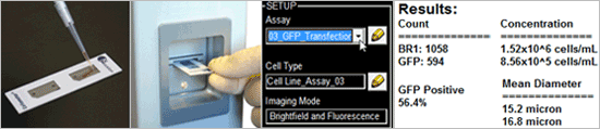
Quantifying GFP Efficiency in 4 Easy Steps
- Pipette 20 µl of cell sample into a disposable counting slide
- Insert slide into the instrument
- Select assay from a drop-down menu
- Click count, acquire image and view cell count, concentration, diameter and percent of GFP positive cells


Leave A Comment