This presentation will focus on using Cellometer image cytometry to effectively determine GFP or other fluorescent protein transduction/transfection efficiency and cell concentration, as well as detect and quantify dual expression in a cell population.
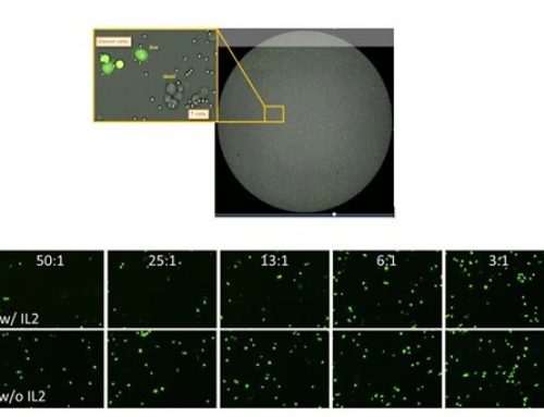
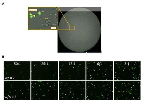
Quantifying GFP can be done in four easy steps. First pipette 20 ul of target cell sample into a disposable counting chamber. Then insert the slide into the instrument, select your assay from the drop-down menu, focus and click count. Within seconds the instrument acquires images, identifies cells with and without fluorescence and automatically tabulates the results. The results include the number of cells counted in bright-field and those that are GFP positive, the concentration for each population, as well as their mean diameter. It also reports the percent of GFP positive cells.


Leave A Comment