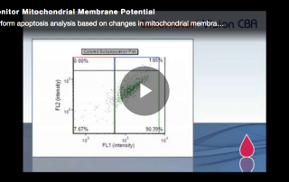
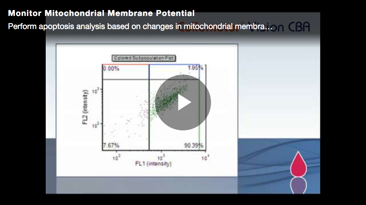
How to Assess Mitochondrial Membrane Potential
This video will show you how to perform apoptosis analysis based on changes in mitochondrial membrane potential with only 20µl of sample.
This video will show you how to perform apoptosis analysis based on changes in mitochondrial membrane potential with only 20µl of sample.
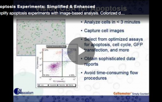
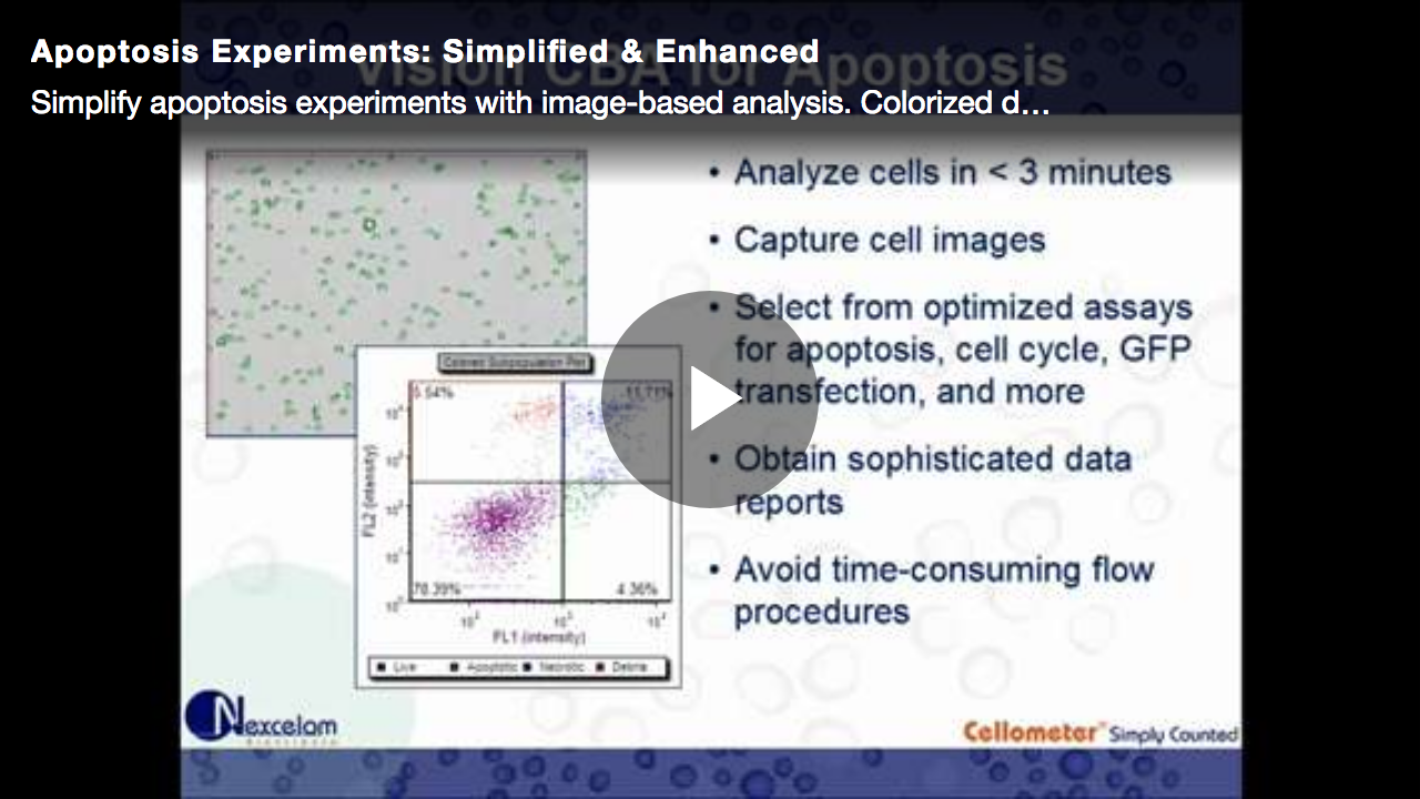
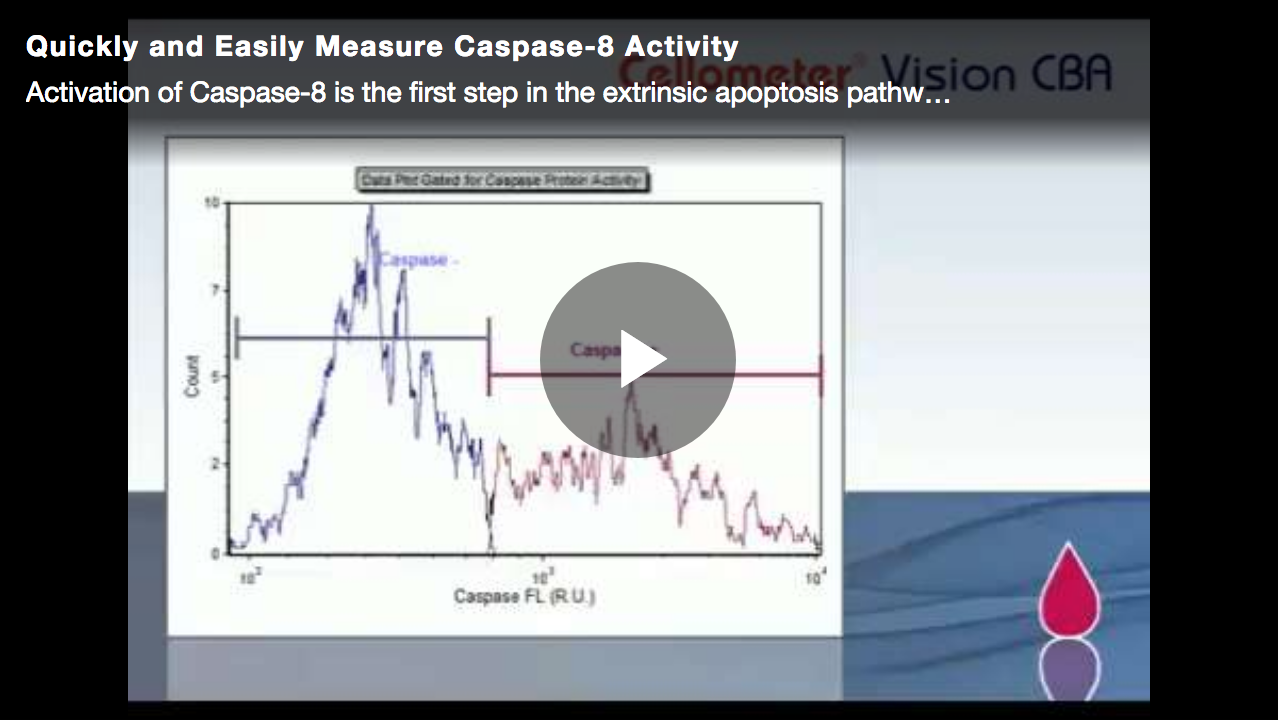
This video will show you how to simplify apoptosis experiments with image-based analysis. Colorized data plots easily identify apoptotic and necrotic cells.
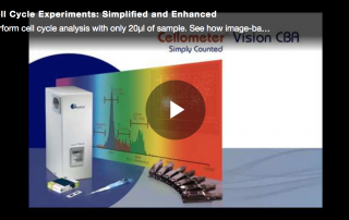
This video will show you how to perform cell cycle analysis with only 20µl of sample. See how image-based analysis simplifies cell-based assays.
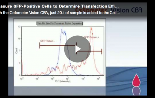
This video will show you how to perform a fast, simple, automated method of GFP analysis that requires only 20 µl of cell sample.
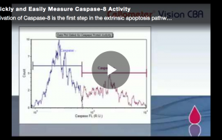
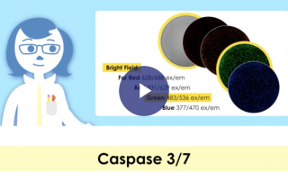
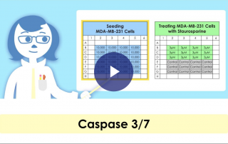
This video will show you how to perform a kinetic apoptosis assay on the Celigo image cytometer using caspase 3/7 detection reagent and suspension Jurkat cells.
This video will show you how to perform a kinetic apoptosis assay on the Celigo image cytometer using caspase 3/7 detection reagent and suspension Jurkat cells.
This video will show you how to perform a kinetic apoptosis assay on the Celigo image cytometer using caspase 3/7 and Hoechst reagents
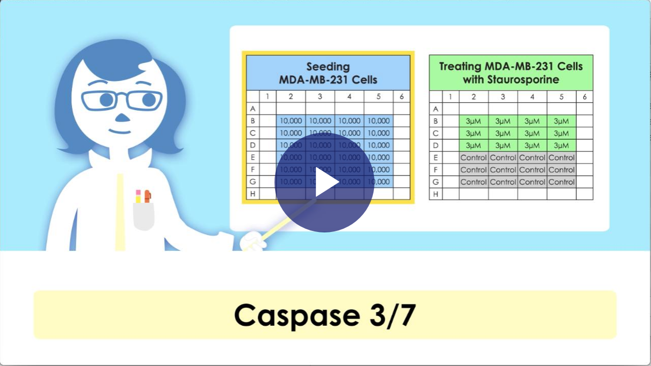
This video will show you how to perform an apoptosis assay on the Celigo image cytometer using caspase 3/7 and Hoechst reagents.
Video: Image autofocus, five-color acquisition, imaging & analysis of every cell to the well edge, fast image scanning & analysis, export data & images, and perform population histogram and 2D gating analysis.
Video: Perform qualitative and quantitative analysis for ADCC, CDC, and cell-mediated cytotoxicity assays using a single platform.
