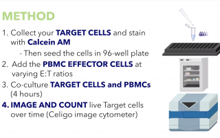
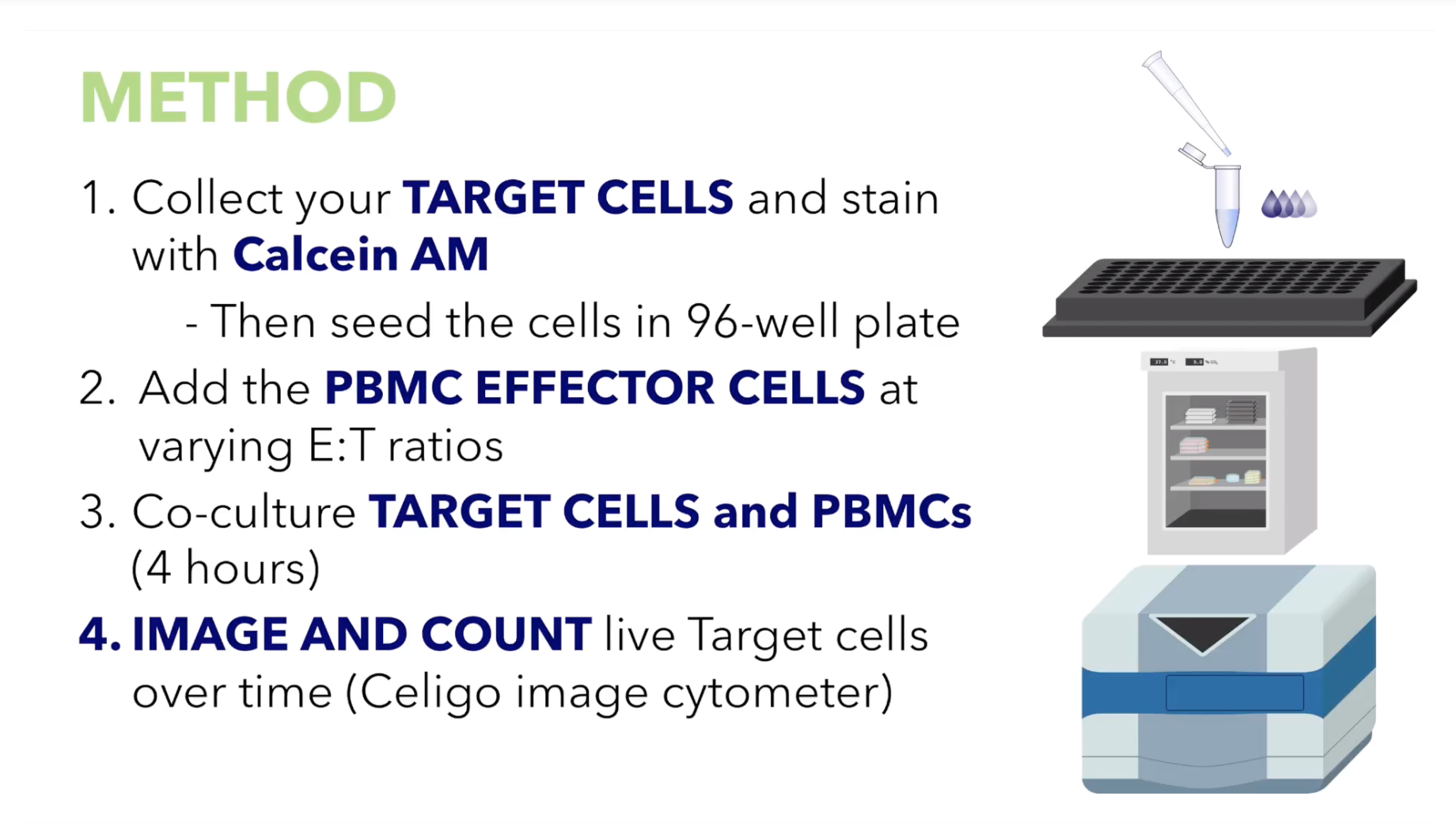
PBMC Mediated Cytotoxicity Using Celigo Image Cytometry
Video: PBMC Mediated Cytotoxicity Using Celigo Image Cytometry. Avoid hazardous radiation in chromium release assays by using image cytometry.
Video: PBMC Mediated Cytotoxicity Using Celigo Image Cytometry. Avoid hazardous radiation in chromium release assays by using image cytometry.
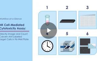
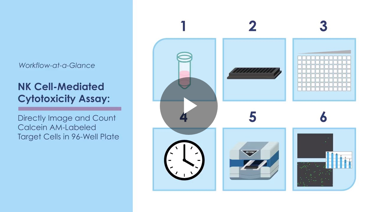
Video: Directly image and count calcein AM-labeled target cells in a 96-well plate using Celigo image cytometer. Concurrent imaging and analysis in 8 minutes.
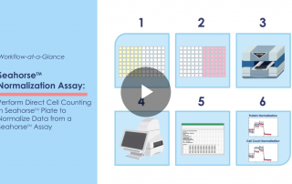
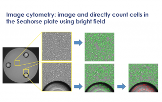
Video: Celigo workflow at a glance to perform direct cell counting or confluence of cells in a Seahorse plate to normalize the data from the Seahorse assay.
Video: How to use Celigo image cytometry to normalize Seahorse™ XF data and eliminate time-consuming protein-based process.
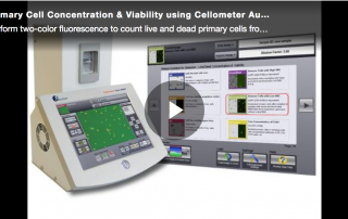
This video shows how to use two-color fluorescence to count live and dead primary cells. PBMCs, stem cells, splenocytes, tumor lysates, and nucleated cells in samples containing debris and red blood cells in 30 seconds.
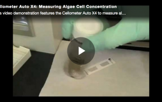
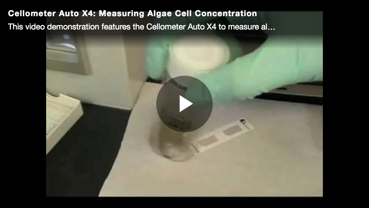
This video demonstration features the Cellometer Auto X4 to measure algae cell concentration from a pond water sample.
This video demonstration explains how to perform 1-step yeast concentration and viability using image cytometry.
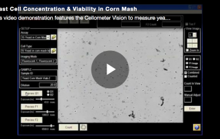
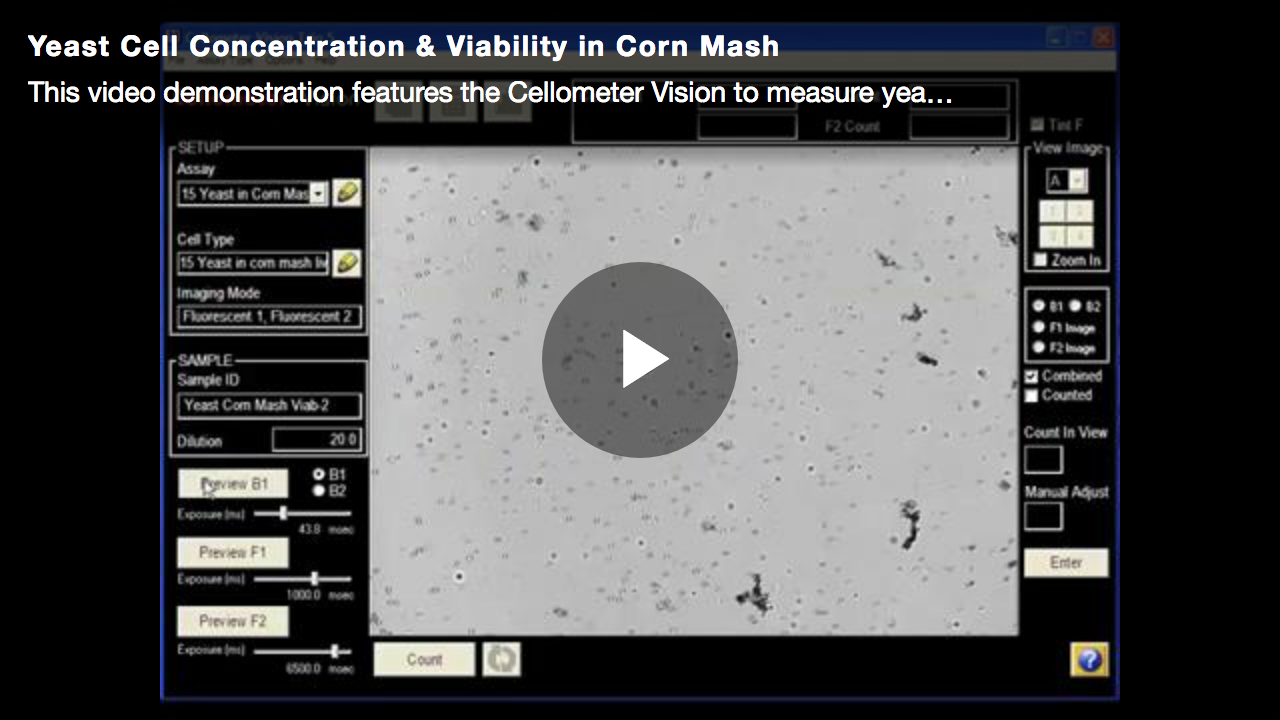
This video demonstration features the Cellometer Vision to measure yeast cell concentration and viability in a highly viscous corn mash sample.
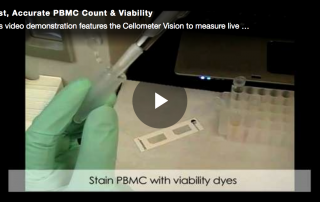
This video demonstration features the Cellometer Vision to measure live cell concentration and viability of human PBMC.
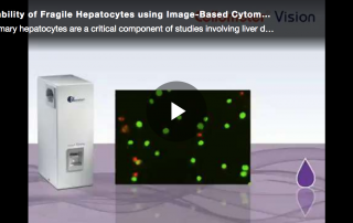
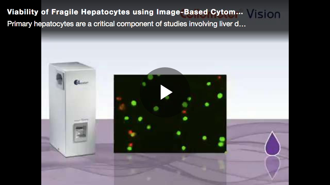
This video will show you how to use bright field and dual-fluorescence imaging to accurately analyze live and dead primary hepatocytes in heterogeneous samples.