What is gene therapy?
Genetic information can vary drastically from cells to cells and the mutations in their gene expressions can disrupt normal cell function to cause cell death or abnormal proliferation, which can lead to a variety of clinical diseases. The first molecular biology techniques were developed in the 1970s to understand gene expression, regulation, and cell cycles. As the technology developed over the years, gene therapy has been employed to investigate the negative or non-existent effects of gene expression on disease-causing cellular functions. Gene therapy is currently utilizing three main approaches to modify the intrinsic expression of certain genes to treat disease:
- Replacing the defective gene with a functioning variant
- Inactivating or suppressing the mutated gene to prevent further damage
- Introducing a new gene to enhance the expression of a targeted gene
When developing cell-based assays for gene therapy, what are the critical points to consider?
Many factors and considerations must be optimized to develop the appropriate cell-based assays for gene therapy development. For example, a kinetic-based assay can determine the proper time to deliver new gene expression and measure the effects over time. Additionally, appropriate target cells and assay conditions should be selected to optimize the assays. Finally, a critical investigation should be conducted to screen patient samples for pre-existing neutralizing antibodies (nAbs) to ensure proper therapeutic results.
Image cytometry-based assays can save time and improve gene therapy development efficiency
Cell-based assays performed with image cytometry support gene therapy research from discovery to development. Nexcelom’s image cytometry and automated cell counting systems support three important aspects of viral vector-based gene therapy research and development.
- Capture and analyze brightfield and fluorescent images for a variety of cell-based assays ranging from cell counting to transduction efficiency detection to cytotoxicity assays
- Perform rapid cell counting and accurately assess cell proliferation and viability of target cell culture
- Conduct high-throughput screening of patient samples for pre-existing neutralizing antibodies
How can image cytometry support the development of gene therapy?
For gene therapy research and development, many cell-based assays are performed requiring optimizations of host-cell quality, quantity, viability, passage number, and cell preparation procedures to ensure consistent and comparable results. Image cytometry systems can optimize these conditions for consistent target cell preparation, determination of the optimal AAV vector, as well as screening for neutralizing antibodies.
- Quality control for high-quality cell culture development
- Rapid cell counting
- Accurate assessment of proliferation and viability
- Optimization of cell culture conditions
- Effortless cloning selection and verification
- Selecting and fine-tuning an appropriate AAV vector
- Simultaneously assess transduction efficiencies and expression levels
- Developing screening assays to identify pre-existing antibodies in patients
- Screen patients for vector-neutralizing antibodies
- Cell-based in vitro neutralization assays
Example: Whole-well imaging enables the identification of non-uniform cell seeding
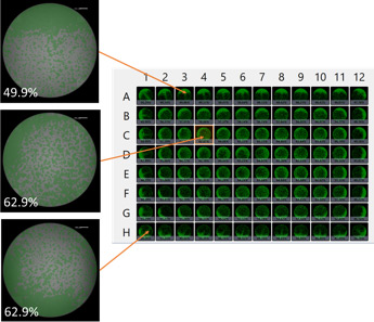
Whole-plate view showing cell confluence for each well. Cell seeding uniformity can vary at different locations within the plate.
These examples of whole-plate images, taken on the Celigo Image Cytometer, indicate cell confluence with a pseudo-color green and show noticeable variations of label-free confluence measurements among wells. Understanding cell growth kinetics and intraplate differences aids researchers in improving data reliability and minimizing variability within experiments. These images underscore the importance of imaging the entirety of every cell, in every well for the whole plate, as this impacts the data and overall quality of results.
Image cytometry technologies save valuable time and resources with the ability to simultaneously count the total number of cells while automatically determining the number of cells expressing targeted antibodies or fluorescent protein-labeled receptors for critical gene therapies. Image cytometry offers rapid screening and monitoring solutions for cell count, viability, apoptosis data, proliferation, cytotoxicity, measuring transduction efficiencies, and gene expression levels to improve the effectiveness of therapeutic development from initial cell line selection to final product quality control.
The image cytometry systems from Nexcelom Bioscience provide user-friendly solutions for cell counting as well as other biological phenomena to support researchers focused on developing new gene therapies. Please visit the gene therapy section of the website to learn more about how image cytometry can support your research and development.


Leave A Comment