Is your 2D assay giving superficial information?
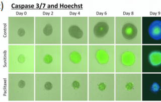
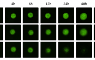
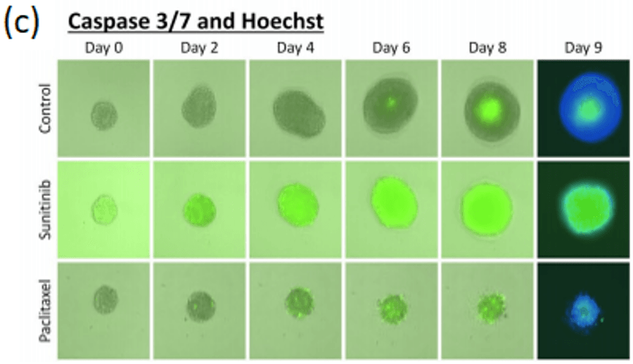
Perform simultaneous imaging and analysis to rapidly capture and quantify spheroid images in every well with advanced software segmentation.
Perform simultaneous imaging and analysis to rapidly capture and quantify spheroid images in every well with advanced software segmentation.
Researchers highlight the need for large-scale screening of cytotoxic cells and their ability to kill target cell lines in both a 2D and 3D fashion.
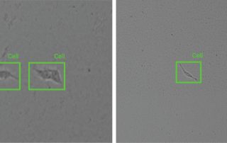
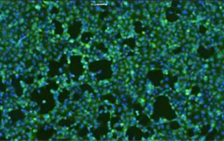
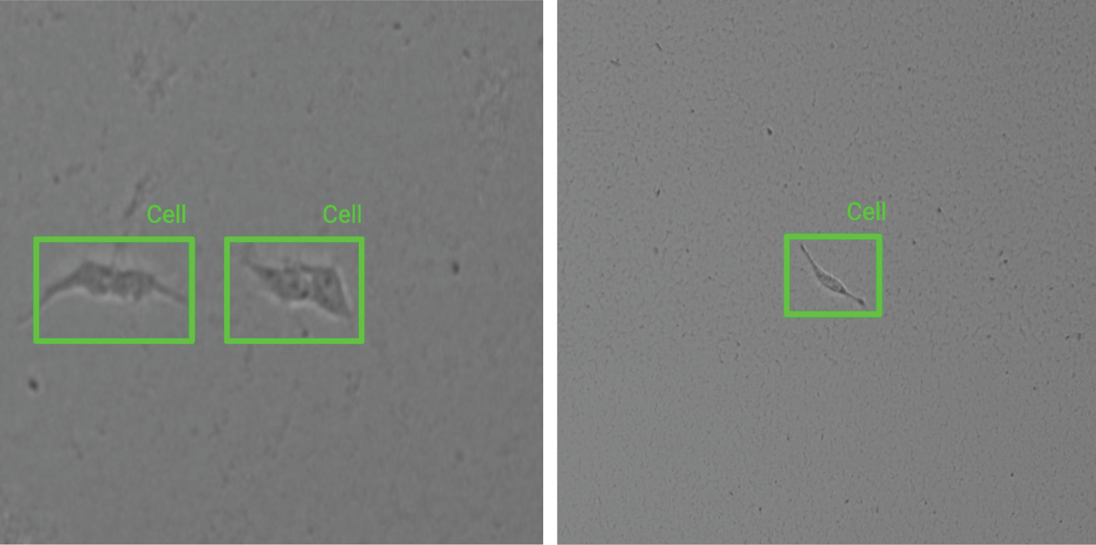
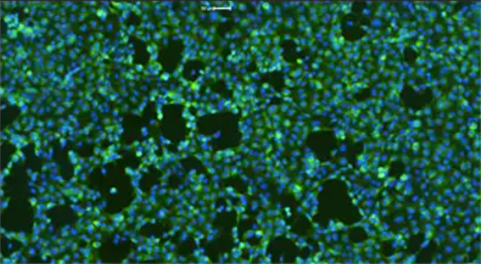
Monoclonal cell culture is critical for many research areas however, the process to establish monoclonal cell lines is time-consuming, and many methods used to monitor monoclonality of cell culture still rely on manual and low-throughput methods.
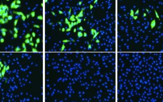
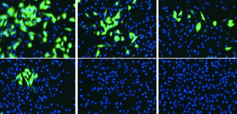
Many leading labs in the field of virology have used the Celigo Image Cytometer to directly measure multiplex cell-based assays in real-time.
Watch the demo on demand: Modern Cell-based Assays for Cell and Gene Therapy Using Image Cytometry
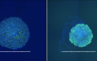
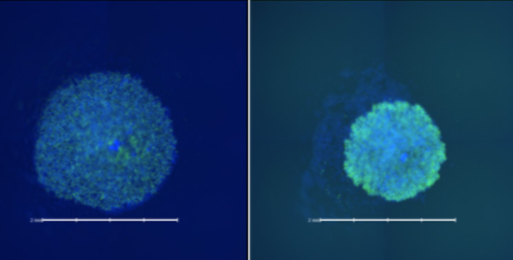
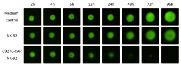
Detection of novel CD276-CAR NK-92 mediated cytotoxicity of neuroblastoma tumor spheroids using plate-based image cytometry
Drug repurposing, also known as drug repositioning is a powerful tool for the rapid expansion of available therapy options by identifying new therapeutic uses from pre-existing drugs.
The Celigo Image Cytometer reduces assay time while providing richer, quantitative readouts to accelerate assessment of vaccine and antiviral compound candidates.
Many factors and considerations must be optimized to develop the appropriate cell-based assays for gene therapy development.
Fast track vaccine development process by conducting assays in 96- or 384-well plates based on enumeration of foci, plaque or single infected cells.
