Impact of Specific Long-term Virus Infections on Host Cells
Viruses are known to cause a wide range of physiological effects on the cell. From changes in gene expression to changes in morphology, some families of viruses can cause persistent infections resulting in long-term infection to the host cell. A clear example of a virus that falls in this category is the varicella-zoster virus (VZV), the causative agent of chickenpox. People who experienced chickenpox at some point in their life and recovered can develop shingles (herpes zoster) later in their life as VZV remains dormant in the nervous system.
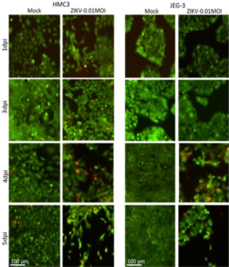
Figure 1. ZIKV infection shown inducting CPE in HMC3 and JEG-3 cells up to five days post-infection. The red stain indicates unhealthy/dying cells and the green stain represents healthy cells.
Other families of viruses can cause a lytic infection or a cytopathic effect (CPE) on the cellular host where the fate of the cell is often death, releasing viral particles to infect neighboring cells for propagation. As more viruses are researched, studies show that cell death is not the only cytopathic effect viruses can elicit on the cell. For instance, the common respiratory syncytial virus (RSV) causes mild cold-like symptoms and secretion of the F protein, inducing the fusion of multiple infected nucleated cells. This behavior is called syncytia.
Understanding Innate Immune Responses through Observation of Virus and Host Communication
Virology researchers use CPE effects pathogens elicit on the host to study host-pathogen interactions. In many cases, this leads to the understanding of innate immune responses or the pathogen’s strategy of immune evasion. Typically, when researchers are studying the relationship of the virus with the host the CPE effects that are typically observed are:
- Cell death
- Cell morphology
- Growth kinetics
- Changes in gene expression or protein levels
- Cytokine production
In many cases, the infection leads to changes in the biology of the cell that leads to beneficial or detrimental effects to the host. This is why understanding how CPE effects manifest in eukaryotic cells is important to study the mechanism of infection of the virus. For example, cells that have survived a viral infection may have an altered cytokine production becoming more resistant to a second infection to the virus. While another class of cells may devote itself to the regeneration and healing of tissue the virus has damaged. Cells that are not able to survive a viral infection are susceptible and are also of particular interest to scientists. This becomes a cell line that can be used to examine how effective the virus is at infecting the cell in the presence of an antiviral compound by scoring CPE effects known to be manifested in this type of cell.
A powerful tool for rapid screening of multiple experimental conditions
The SARS-CoV-2 pandemic has placed a critical focus on viral research and the Celigo image cytometer has played a vital role in virology development. The Celigo is a powerful tool with user-friendly software providing a variety of applications including screening CPE effects and providing cell morphology and cell death data.
Types of CPE effects that can be monitored on the Celigo:
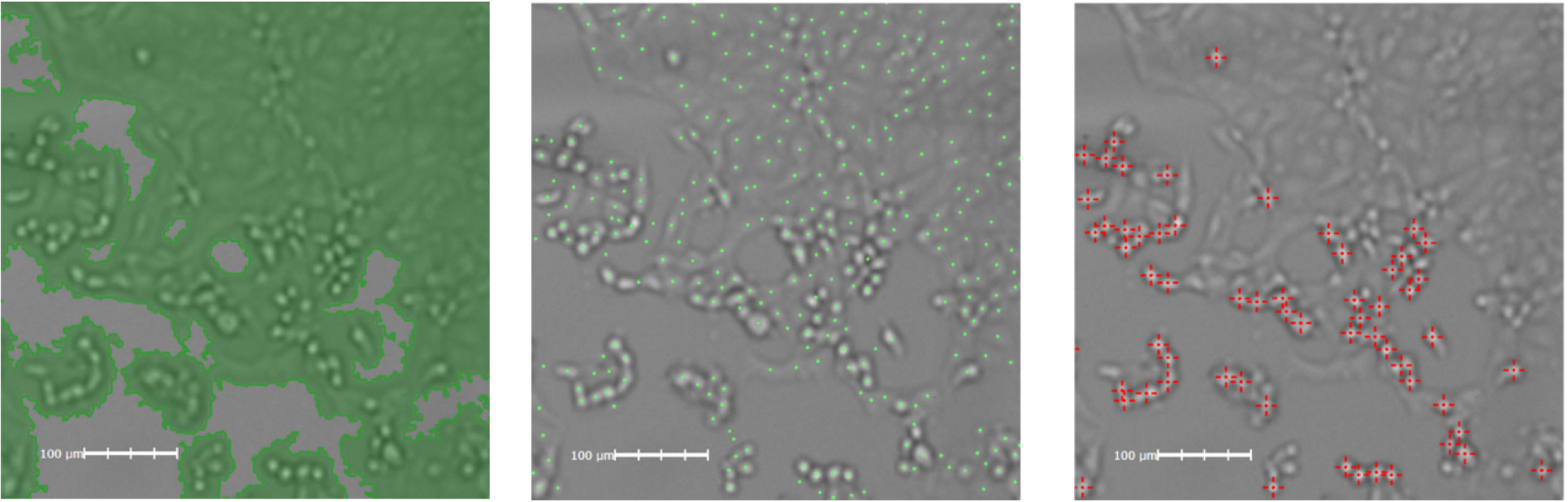
Figure 2. Loss of cell density measured as measured in the Celigo image cytometer. Confluency application (left), Direct cell count (center), Cell morphology (right)

Figure 3. Workflow from scanning a plate to image analysis complete with gating can take 6 min for a 96 well plate.
Incorporating the Celigo Image Cytometer in the experimental workflow can be a time saver. For instance, various types of vessels from 6 well to 1536 well plate with adherent or suspension cells can be loaded manually in the Celigo for analysis. Additionally, the process can be fully automated by adding a plate stacker for a more efficient workflow and high-throughput projects. The F-theta lens allows for whole well, high-quality imaging at 1 µm/pixel resolution. These unique scanning mirrors allow for the fast scanning of the whole plate without compromising the integrity of the data. Finally, the Celigo software includes a comprehensive suite of image analysis algorithms and gating capabilities to analyze the data in real-time and the software automatically saves all the data in a SQL database, making it easier to log, access, and re-analyze previous data.
Aid in the Rapid Screening of Effective Therapies Against New and Emerging Pathogens
Classic experiments like plaque and microneutralization assays can more rapidly derive biological information when coupled with image cytometry. Screen thousands of drugs and neutralizing antibodies in a fraction of the time. The Celigo Image Cytometer allows the imaging of infected cells at 1 µm/pixel resolution in a variety of plate formats which allows for numerous experimental permutations. Researchers also have access to Nexcelom’s cell counting expertise to assist in the ease of developing fit-for-purpose methods and nurturing collaborations to achieve results more efficiently.
Learn more about modern virology assays.
References:
- Heaton, N., Revisiting the concept of a cytopathic viral infection, PLoS Pathog, 2017, 13(7)
- Amanat, F., An in vitro microneutralization assay for SARS-CoV-2 serology and drug screening, Current Protocols, 2020, 50
- Martinez Viedma, M, et.al., characterizing the different effects of Zika virus infection in the placenta and microglia cells, Viruses, 2018, 10(11):649


Leave A Comment